Different bacteria produce different “effects” in broth cultures. For example, sedimentation could occur, slime production, surface layers produced on the broth, etc. Certain bacterium will grow more effectively on broth cultures.
Despite bacteria being incredibly versatile creatures, all bacteria have optimum conditions, and conditions which will harm them. Temperature is one of the most important factors due to enzymes within the bacterial cell. Bacteria often have thousands of different types of enzymes in their cells, which up to a certain temperature will increase in efficiency and productivity of the bacterium, i.e. reproduction will occur faster. This is because as temperature increases, the kinetic energy of the enzyme and the substrate will increase, i.e. they move around faster. This increases the probability of an enzyme and its substrate colliding, and colliding in the right orientation, with enough energy for a reaction to occur, i.e. an increase in temperature increases the probability of a reaction occurring, therefore meaning metabolic activity and eventually growth is increased. However after the optimum temperature, the rate of the reaction decreases, because the temperature begins to damage to the bonds, e.g. disulphide bridges, within the enzyme. This changes the shape of the active site of the active site on the enzyme, meaning that the substrate can no longer fit into the site and bonds cannot be broken/made. Therefore growth of the colony will decrease in rate or cease. Other conditions also effect bacterial growth, e.g. Oxygen concentration, pH, presence of moisture.
Examples of species of bacteria include Escherichia coli (E.Coli), and Micrococcus Leteus (M.Leteus). M. Leteus. Closely related to M.lylae, is a gram positive cocci. Classed as an opportunistic pathogen, it is commonly found on human skin, causing septic shock and pneumonia, but is not found on animal skin and cannot survive in soil. Their ideal temperature for growth is 37 degrees Celsius. When cultured on solid medium, it is known to produce cream white circular colonies.
E.Coli bacteria, normally found in human stool, can exist as a non harmful strain or as a harmful strain, most commonly strain 157. Diseases of E.Coli include diarrhoea, which can be treated with anti-biotic, and meningitis. When grown on solid plates, E.Coli produces small yellow circular colonies.
(, 2004) (, 30/01/2004) (Prescott et al, 2002)
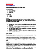
~ Method
The method used was the same as the method described in the course module guide. The first method of streaking was as below.
The second technique is shown below:
The only unique thing was the style of streaking used for the last nutrient agar plate. For this plate, the “smiley face” technique was adopted to try and separate out individual colonies for the mixed culture. The method for this highly skilful technique is outlined below;
-
Using the aseptic technique (described in the introduction section), the inoculating loop was placed approximately one third of the way down the plate, and approximately one third inward, and streaked as indicated on the diagram (below).
- This was done again on the opposite side of the plate, giving an “eye effect”.
- A similar sized streak was placed on the centre of the plate, giving a “nose” effect.
- A roughly semi-circular final streak was applied near the bottom of the place, approximately in the middle.
- Diagram shown below:
- An effect known as “hair” can also be adopted but this technique was considered too advanced for this practical.
~ Results
See attached sheets of plain paper.
~ Discussion & Conclusions
All the controls used for this experiment (the agar plate and the nutrient broth), were both completely clear and absent of any obvious bacterial growth and colonisation, suggesting that both the agar plates and the broth were clear of bacteria before the experiment was started, and proving the only bacteria present was that added by us, i.e. M.Leteus and E.Coli and anything present in the soil.
The 3 agar pates left in the open air had very little bacterial growth on them at all, and very little variation in what growth there was. The plate incubated at room temperature had only two obvious areas of colonisation, both of which were apparently differently different species. A small, white asterix shaped colony grew near the centre of the plate, and yellow, circular colony approximately 1.5mm in diameter. This is the only plate which the white, asterix shaped colony appeared on, therefore it is impossible to compare growing conditions i.e. temperature in this case. The only conclusion that could possibly be drawn is that this particular bacterium’s optimum growing temperature is room temperature, i.e. approximately 25 degrees Celsius. This particular bacterium could be isolated and grown, and further experimentation could be done to find this bacterium’s optimum temperature and other things.
The plate incubated at 30 degrees Celsius was colonised by the most bacteria and by the largest variety of bacteria, suggesting that this is the optimum temperature of incubation for most of the air born bacteria present in the air surrounding the plate. The majority of the colonies were small, yellow, circular colonies, 1-4mm in diameter. On the room temperature plate there was one yellow colony of similar size, and on the 37 degree Celsius plate there was again one yellow colony of similar size. It is highly likely that these three colonies are in no way related and are of completely different species, but comparative purposes I will assume that they are of the same species. The plates suggest that 30 degrees Celsius is the optimum temperature for this bacterium because the most growth occurred on this plate. One other colony was present. This was a relatively large colony, not circular, and was slightly textured on the surface. This bacterium was only found on this plate and therefore the only conclusion that could possibly be drawn is that its optimum temperature is 30 degrees Celsius also.
As mentioned in the last paragraph only one yellow colony was present on the 37 degree plate, and the agar was mostly colonised by white circular colonies, approximately 1 mm in diameter. If we again assume that the white colonies were all of the same species, we can conclude that the optimum temperature for this bacterium’s growth is 37 degrees.
The remaining 3 agar plates were all incubated at the same temperature for the same amount of time, therefore the only variable which could have effected the separation of different colonies was the streaking method. The first streaking method adopted was the line method (see attached sheet 2, diagram 6).
The smiley face method adopted for the 3rd agar plate was not particularly effective at separating colonies. While a few creamy white colonies were distinguishable from the solid streaks, in the pattern known as “spots”, the previous two methods were far more effective. Both the line and the snake methods were quite effective at separating out the E.Coli and the M.Leteus colonies. I believe the creamy white colonies to be M.Leteus, and I think that the line method is best at isolating this bacteria. I believe the snake method to be best at isolating the yellow colonies which I believe to be E.Coli.
The liquid culture for M.Leteus was fairly clear before and after swirling. Before swirling the bacterium colonised the bottom of the glass container. Because of this, and because of a, much larger presence of M.Leteus on the agar, I conclude from this that M.Leteus is best adapted/”likes” solid media for growth rather that liquid media.
The E.Coli was cloudy before and after swirling and produced slime which did not disperse after swirling. This seemed grow well in the liquid culture compared to M.Leteus and grew well on the solid agar, suggesting that it may be more versatile than M.Leteus.
The soil sampled produced slime which was present before and after swirling. It also produced a surface layer before and after, and was cloudy before and after. I conclude from this that because of all the different things present, e.g. slime, surface layer, cloudiness, that more than one species of bacterium was present.
~ Bibliography
-
, 30/01/2004
-
, 2004
-
Prescott, Harley & Klein, (2002), Microbiology, 5th ed. McGraw Hill.