The Geiger counter is one of the oldest and simplest of the many particle detectors. The counter was developed in the early part of the twentieth century by Hans Geiger and Wilhelm Müller, shortly after the discovery of radioactivity. A wire electrode runs along the centerline of a cylinder with conducting walls. The tube is usually filled with a monatomic gas such as argon at a pressure of about 0.1 atmospheres. A high voltage, slightly less than that required to produce a discharge in the gas, is applied between the walls and the central electrode. A rapidly moving charged particle which gets into the tube will ionize some of the gas molecules in the tube, triggering a discharge. The result of each ionizing event is an electrical pulse that can be amplified to activate earphones or a loud speaker, making the counter useful in searches for radioactive minerals or in surveys to check for radioactive contamination.
Scintillation counters are made from materials which emit light when charged particles move through them. To detect these events and to gain information about the radiation, some means of detecting the light must be used. One of the first scintillation detectors was a glass screen coated with zinc sulfide. This sort of detector was used by Ernest Rutherford in the early versions of his classic experiment in which he discovered the nucleus of the atom by scattering alpha particles from heavy atoms such as gold. The scattered alpha particles hit the scintillating screen, and the small flashes produced were observed by experimenters using only the human eye.
1: terrestrial: originated from the planet earth (not alien).
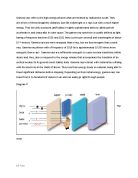
Diagram02
Gamma rays are used in medicine, the nuclear power industry, the military, scientific research, industry, and various consumer products.
Gamma radiation is also important in the medical sciences. Ionizing radiation is more harmful to cells when they are dividing (replicating their DNA), and this has led to the use of gamma radiation for the treatment of cancer. The rapidly dividing cancer cells are more susceptible to the radiation than the healthy cells and are destroyed selectively without the need for surgery. Molecules that contain very short-lived isotopes are used as tracers to study the distribution of the molecules within the body. Due to the sensitivity with which radiation is detected, only minute quantities of the chemical are required. This information is used for both diagnostic and research purposes. For example, it has enabled the three dimensional imaging of the brain as a function of particular stimuli.
Diagram 03
Military uses of materials and processes that emit X-radiation and gamma radiation include the production of materials for nuclear weapons and the testing and use of nuclear weapons. In 1945, atomic bombs were detonated over Hiroshima and Nagasaki, Japan. Between 1945 and 1980, nuclear weapons were tested in the atmosphere of the Northern Hemisphere; during the most intense period of testing, from 1952 to 1962, about 520 tests were carried out.
Diagram 04
Several industrial processes use ionizing radiation. Industrial radiography uses gamma radiation to examine welded joints in structures. In the oil industry, gamma radiation or neutron sources are used to determine the geological structures in a bore hole (a process called “well logging”). Ionizing radiation is also used to sterilize products and irradiate foods (to kill bacteria and parasites).
03 retrieved from:
04 retrieved from: http://www.insidestory.iop.org/radio.html
Biological damage by ionizing radiation is related to dose and dose rate, which may affect the probability that cancer will occur. Radiation dose is a measure of the amount of energy deposited per unit mass of tissue and may be expressed as the absorbed dose, equivalent dose, or effective dose. The standard unit for absorbed dose is the gray, which is equal to 1 J/kg of deposited energy. The absorbed dose formerly was expressed in rads (1 Gy = 100 rads). The biological effect of high-LET radiation is greater than that of low-LET radiation at the same absorbed dose; therefore, a dose measurement independent of radiation type was derived to reflect the biological effectiveness of radiation in causing tissue damage.
Diagram05
05 diagram showing the effect of gamma rays on tissues. Retrieved from: http://www.nidcd.nih.gov/news/releases/09/pages/06_24_09.aspx
Resources:
Report on Carcinogens, Twelfth Edition (2011) retrieved from :
Gamma history 2011 retrieved from : http://infotrac.galegroup.edu /itweb/tlemea_scirc?cause=http%3A%2F%2Fgalenet.galegroup.com%2Fservlet%2FSciRC%3FlocID%3Dtlemea_scirc%26bi%3DSU%26bt%3Dgamma%2Brays%26c%3D5%26t%3D1%26ste%3D21%26docNum%3DCV2432500315%26st%3Db%26tc%3D31%26tf%3D0&cont=&sev=temp&type=session&sserv=no
NDT resource center on ionizing radiation retrieved from:
G-nelson and E riley on gamma rad. Retrieved from:
High energy radiation from black holes retrieved from: http://press.princeton.edu/titles/9092.html