What causes cystic fibrosis?
What causes cystic fibrosis?
Cystic fibrosis (CF) is an autosomal recessive, hereditary disease, affecting 7500 people in the UK (Cystic fibrosis trust UK). The first documented case in 1936 described the condition as fibrocystatic disease of the pancreas. Before the 1960's the average life expectancy was five years; today it stands at around 31 and is expected to grow to around 40 years in the next decade as a result of advances in treatment methods. Every week five babies are born with CF and three people a week die from it usually as a result of lung damage (Cystic fibrosis trust UK). The common feature for affected systems is epithelial layers that secrete a mucus layer; including: the respiratory system, gastro-intestinal tract (including the pancreas) and the genito-urinary tract. Most complications associated with the disease arise from infection of the airways or other organs by a number of pathogenic bacteria, most notably Pseudomonas aeruginosa. Other bacteria and/or viruses may colonise mucosal surfaces and lead to infection or pave the way for more serious infections.
CF is caused by a mutation, which occurs in the Cystic Fibrosis Transmembrane conductance Regulator (CFTR) gene. The most common mutation gives rise to a protein missing amino acid 508, termed ?F508 CFTR, and accounts for around 70% of the cases reported in the UK. Patients homozygous for ?F508 generally have severe pulmonary complications and pancreatic insufficiency. Around 400 other mutations in this gene have been documented and this is most probably related to its size - 250 kbp - and large number of exons. The various phenotypes in cystic fibrosis relate to the different genotypes possible ie. the mutation may result in reduced abundance or function of CFTR within the cell or the protein could be completely absent. Due to the prevalence of the ?F508 mutation and its almost complete fatality rate, this essay will concentrate on the implications and possible solutions? to it.
In order to understand the causes of cystic fibrosis, it is necessary first to explain the way in which the normal protein works. The CFTR gene may perform a number of functions that have yet to be elucidated, but, a number have been ascertained so far, largely as a result of the cloning and sequencing of the gene itself. Now its structure is known scientists have confirmed the CFTR protein to be a channel for Cl- and HCO3- ions that is present in a number of organs.
The wild type CFTR gene produces a molecule that becomes embedded in the cell membrane and consists of two halves forming a channel, each side having a nucleotide binding fold capable of binding ATP. In the presence of adenylate cyclase the bound ATP can be converted into cAMP. The symmetrical halves are joined by an R-domain, which is phosphorylated by protein kinases in the; when this happens a conformational change causes the pore to open as the R-domain moves away from the pore aperture. These protein kinases are activated by cAMP so when ATP and adenylate cyclase are ...
This is a preview of the whole essay
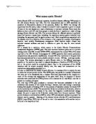
The wild type CFTR gene produces a molecule that becomes embedded in the cell membrane and consists of two halves forming a channel, each side having a nucleotide binding fold capable of binding ATP. In the presence of adenylate cyclase the bound ATP can be converted into cAMP. The symmetrical halves are joined by an R-domain, which is phosphorylated by protein kinases in the; when this happens a conformational change causes the pore to open as the R-domain moves away from the pore aperture. These protein kinases are activated by cAMP so when ATP and adenylate cyclase are present the channel may be opened. . Opening of the channel allows chloride (Cl-) and bicarbonate (HCO3-) ions (Wine, 2001) out of the cell or into it (in association with another ion) or both, as in the respiratory tract.
Figure 1 - Proposed structure for CFTR when incorporated into the cell membrane (adapted from Cystic fibrosis - AUTHOR)
The CFTR protein acts only as an ion channel and therefore must come under the influence of some regulatory elements that affect levels of cAMP and/or protein kinases. Cells expressing the CFTR may be influenced by paracrine and/or endocrine cells whose hormones or factors may induce/inhibit the production of cAMP thus bringing about the opening/closing of the channel. cAMP is often used as a second messenger, being activated when hormones bind to a cells membrane, the molecule then activates protein kinases as described.
SCHEME FOR ACTIVATION BY cAMP
The various types of tissues expressing the CFTR perform many different functions and thus have different uses for the protein. The CFTR plays a crucial role in regulating ion levels in pancreatic secretions. Acini cells in the pancreas produce enzymes and peptides for secretion into the duodenum? along with bicarbonate and phosphate ions that act as buffers, neutralising the acids present in chyme from the stomach. In CF patients pancreatic complications usually involves the accumulation of secretory material, blocking the ducts and leading to tissue damage. Affected cells release lytic enzymes that damage surrounding cells leading to loss of function, fibrosis and fatty infiltration.
The respiratory tract is lined with epithelial and goblet cells that secrete mucus containing ions, peptides and immunoglobulins. The mucus forms a protective covering for cells acting as a barrier to dust, allergens and pathogens that would otherwise irritate the sensitive epithelium. The main constituent of mucus is a glycoprotein called mucin that has many carbohydrate side chains (usually sialic acid) and these molecules interlock to yield a fluid, elastic layer. Cilia lining the airways beat continuously producing a current that moves mucus (up or down) towards the oesophagus where it is swallowed and moves to the stomach. The mucus and anything held in it (including any dangerous material) is broken down by digestive juices and enymes; the acidic nature of the stomach is sufficient to destroy most disease causing micro-organisms. The mucus layer must be maintained within relatively constant parameters e.g. solutes dissolved in it affect viscosity and are regulated so that the cilia are able to continue moving mucus along the airways.
When molecules of mucin are released onto the epithelial surface the negatively charged side chains attract water molecules and thus become hydrated. If mucin becomes over-hydrated the cilia will not be able to move mucus along the airways. Similarly if mucin becomes dehydrated the mucus will become too thick for cilia to move, the latter (called the thick mucus hypothesis) occurs frequently in cystic fibrosis and leads to complications that are potentially fatal. Epithelial cells in the respiratory tract normally express the CFTR protein, as mentioned earlier this is an ion channel present in epithelial cells and helps to regulate ion concentration in the mucus layer. Cl- and Na+ in mucus act as counter ions, which antagonise the hydration process and are moved in and out of epithelial cells when necessary so that the mucus remains at a manageable viscosity. Epithelial cells lacking the CFTR protein are unable to regulate levels of Cl- and Na+ in the mucus and so most of the water molecules associated with mucin are displaced leading to a thick mucus which the cilia are unable to move.
SEE HANDOUT AND TEXTBOOK
Diagram of Chloride/Sodium transport
Diagram for mucin production
The bicarbonate before chloride hypothesis
Most of the symptoms associated with the CF are thought to stem from cells' inability to transport Cl- ions, however, the CFTR protein has been found to conduct other substances such as HCO3- (Wine, 2001) and more recently reduced glutathione (GSH) to be described later. The former compound forms an important constituent of pancreatic secretions that empty into the duodenal ampulla. HCO3- acts as a buffer to the highly acidic chyme produced in the stomach, thus protecting the enzymes in the pancreatic juices and the cells of the intestine from denaturation or damage. In CF patients, acini cells in the pancreas are deficient for the CFTR gene and as a result are not able to secrete HCO3- into the juices they produce. CF pancreatic secretions will be deficient in the necessary ions as other positively charged ions will not move out of the cells to counter the anion efflux. This has the effect that such secretions will be thicker as water molecules will be less likely to diffuse out of cells in association with the ions, and subsequently the secretory ducts (that empty into the pancreatic duct) become blocked. Blockage of these ducts initiates the process of apoptosis, followed by infiltration of fibrous scar tissue and eventual lack of function in the pancreas. Further, those ducts that remain unblocked for a time continue to secrete a solution rich in enymes but unable to counter the acidic nature of chyme and so the enzymes present become denatured and less able to digest food material. The inability to digest food particles leads to malabsorbtion in the small intestine, and coupled with other adverse effects of the disease causes CF patients to be under nourished despite dietary supplements (Cystic fibrosis - SHALE?).
GSH Transport
The CFTR has been found to transport a number of molecules across cell membranes, e.g. reduced glutathione (GSH), a large organic anion with powerful antioxidant properties. The fact that the CFTR conducts tripeptides as well as small molecules has paved the way for new hypotheses about its role in the cells of the body. It is possible that the CFTR allows the passage of a variety of different molecules that may have wide ranging effects on the pathology of cystic fibrosis. Many of the clinical manifestations of the disease remain unaccounted for by the model proposing a defective Cl- channel as the cause of the disease. Recent hypotheses such as Hudson's (2001) offer alternative viewpoints for considering the CFTR gene, it may be more accurate to see it as a multi-functional channel, performing a range of important regulatory functions.
Normally GSH is synthesised in cells e.g. epithelial cells, leukocytes; and secreted into the extracellular milieu where it can be oxidised by reactive oxygen species (ROS), such as hydrogen peroxide or superoxide, that would otherwise damage the surrounding tissue. Such ROS are common in the airways, which can sometimes have elevated levels of GSH over 100 times the normal level. GSH deficiency is by no means restricted to CF, as it has been recorded in a number of other respiratory illnesses e.g. chronic obstructive pulmonary disease (COPD). The human antioxidant system comprises a number of molecules (some of which are enzymes) that offset the effects of ROS, however, GSH is a precursor for one of these enzymes. GSH also forms an interdependent system with two other antioxidants i.e. when GSH levels fall the other components' levels also fall. The result of lowered GSH in the extracellular milieu therefore leads to a breakdown of the whole system, which would previously have protected the epithelium. However, the complications arising through depletion of GSH levels must not be interpreted as causative of CF, but rather as an exacerbation of symptoms all ready present.