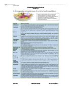
Understanding the basic structure of a cell
Cells Structure
All cells have organelles; these are the specialised structures within a cell that help it to perform the specific functions.
The Nucleic Acids
The nucleic acid consists of two complex chemicals which carry genetic information within the cells. These complex chemicals are called DNA (deoxyribosenucleic acid) and RNA (ribosenucleic acid).
The DNA carries the genetic code and its structure allows it to store information, pass information on to RNA so that proteins can be made, and also to copy itself, this allows the genetic code to be passed into new cells. DNA consists of a double strand of alternating sugar and phosphate molecules, and has four bases; A (adenine), T (thiamine), G (guanine) and C (cytosine). The bases project out from the sugars at right angles. These bases pair up in specific orders, A will always pair with T and G will always pair with C. when these bases are paired, they are held together by hydrogen bonds. The two strands are then twisted together, each base twists and with every ten pairs a full 360 turn has been made.
RNA consists of only one strand, similar to DNA but also has alternating sugar and phosphate molecules. There are two types of nucleic acid; deoxyribonucleic acid (DNA) and ribonucleic acid (RNA). Then there are three different RNA types: messenger (mRNA); transfer (tRNA) and ribosomal (rRNA). RNA bonds the same as DNA except it has no thiamine; it has Uracil (U). The RNA pairs with DNA to produce mRNA; this then travels to the ribosome, which it combines with the amino-acids, which make the proteins required.
DIFFERNENCES BTWEEN DNA and RNA
Sugar-phosphate backbone phosphate
Sugar
Base
Hydrogen bonds
During protein synthesis the part of the DNA that is responsible for the formation of a particular protein (e.g. insulin) uncoils and splits.
Portions of the RNA then join with the DNA where the bases complement each other,
A strand of RNA is then formed, known as messenger RNA (mRNA)
The mRNA leaves the nucleus through the nuclear pores and travels to the ribosome
In the ribosome, there are amino acids (aa),
The mRNA is then combined with its corresponding aa, which then forms the protein (e.g. insulin)

Understanding how substances are transported across the cell membrane
Cell Membranes
The cell surface membrane is the boundary between the cell and its environment; it has very little mechanical strength but plays a vital role in controlling which materials pass into and out of the cell.
The cell membrane is formed from a double layer of phospholipids molecules, arranged tail to tail, the cell membrane is a very complex structure, which is studded with proteins. These can be embedded in the membrane or they can penetrate the bilayer forming ...
This is a preview of the whole essay
Understanding how substances are transported across the cell membrane
Cell Membranes
The cell surface membrane is the boundary between the cell and its environment; it has very little mechanical strength but plays a vital role in controlling which materials pass into and out of the cell.
The cell membrane is formed from a double layer of phospholipids molecules, arranged tail to tail, the cell membrane is a very complex structure, which is studded with proteins. These can be embedded in the membrane or they can penetrate the bilayer forming pores through which molecules can pass.
Cell membranes contain phospholipids, proteins, cholesterol and polysaccharides. The phospholipids are a major constituent of cell membranes. They naturally form membranes in water because they automatically arrange themselves into a bilayer that is practically impermeable to water and anything that is water soluble. The membrane proteins act as hydrophilic pores; these are water filled channels that allow water-soluble chemicals to pass through. These pores are usually small and highly selective, proteins in the membrane that form pores usually span the entire membrane, but other proteins with other functions can occur only in the top or bottom layer of lipids;
Membrane proteins have several functions, they:
- Form pores through which water and water-soluble chemicals can pass
- Act as carriers in active transport
- Form receptor sites for hormones
- Are important to cell recognition
Cell surface membranes can also contain cholesterol molecules, which fit in between the phospholipids.
Polysaccharides, branched polymers of simple sugars, stick out from the outer surface of some membranes like antennae. They attach to lipids to form glycolipids, or to proteins to form glycoproteins. The glycolipids and glycoproteins help cells to recognise other cells, which allow the body to recognise between body cells and invading bacteria.
Materials pass in and out of the cell by the following processes;
Diffusion –passive process that doesn’t require energy from the cell, it is the movement of particles within a gas or liquid from a region of high concentration to a region of lower concentration. Diffusion moves substances over short distances. For example, diffusion occurs within the lung; oxygen diffuses through the thin epithelium of the alveolus and into the blood,(this journey is usually less than a hundredth of a millimetre), it is then carried away to parts of the body by the circulatory system. There are several factors that affect the rate of diffusion these are
- The surface area between the the two regions- the greater the surface area, the greater the rate of diffusion
- Length of diffusion pathway – this is the distance over which the diffusion occurs.
- The concentration gradient – diffusion is more efficient if the concentration gradient can be maintained
- Size and nature of particles to be diffused – fat-soluble substances can diffuse through the lipid bilayer of the membrane. Water soluble substances must pass throught the protein pores, these tend to be small and selective. Generally larger molecules cannot diffuse into or out of the cell at all.
- Temperature – at higher temperatures the molecules have more kinetic energy and diffuse more quickly.
Facilitated diffusion is a passive process. It is the diffusion enhanced by specific proteins in the cell membanes.
Two types of proteins are responsible for facilitated diffusion;
- Carrier proteins – take particular substances from one side of the membrane to the other. As soon as the molecules binds with the carrier protein , the protein changes in shape so that the molecule ends up on the other side of the membrane where it is released.
- Ion channels – proteins that open and close to control the passage of selected charged particles. Proteins with a central hole lined with polar groups, ion channels facilitate the diffusion of charged particles (Ca2+, Na+, Cl- ions). Cells use ion channels to control the movement of ionic substances between themselves and other cells, and also to regulate the ionic composition of their cytoplasm.
Osmosis
Osmosis is the movement of water molecules across the cell membrane, from an area of high concentration to an area of low concentration until an equilibrium is reached. Osmosis is a passive process, so no energy is required for this to happen.
When blood plasma becomes hypertonic to red blood cell cytoplasm, as it does during severe dehydration, water is lost from the red blood cells, which shrink and become crinkled (crenated). At the other extreme, hypotonic plasma causes red blood cells to burst, this process is known as haemolysis – blood splitting.
To protect cells from the effects of osmosis we must maintain body fluids at the same solute concentration as our cell cytoplasm.
Active transport
The movement of molecules across the cell membrane from low to high concentration.
In active transport metabolic energy, which is produced by the cell and stored as ATP (adenosine tri phosphate), is used to drive the transport of molecules and ions across cell membrane. There are differences between active transport and diffusion.
Active transport is similar to facilitated diffusion. Cell membranes contain specific carrier proteins that combine with the substance to be transported, allowing the substance to pass rapidly across the cell membrane.
These processes all involve active transport;
- Absorption of amino acids from the gut into the blood
- Reabsorption of glucose and other useful molecules in the kidney
- Pumping sodium ions out of cells
- The exchange of sodium and potassium ions which allow conduction of the nerve impulses.
Bulk Transport
Bulk Transport transports larger volumes of materials into and out of the cell; this can be liquids or solids. The different processes of bulk transport are called; Endocytosis and Exocytosis.
Endocytosis
This is the process in which a substance gains entry into the cell without passing through the cell membrane.
There are two main types of endocytosis; phagocytosis and pinocytosis.
Phagocytosis – cells takes in (phagocytose) solid particles like bacteria and old red blood cells so they can be destroyed. This is done by the cell’s cytoplasm, which flows round solid material, when the particles are fully surrounded a section of the cell membrane is pinched of to form a vesicle around the particle. The membrane and the solid particles inside the vesicle form a food vacuole. This allows for the lyosomes to fuse with the vacuole membrane, which allows digestive enzymes to enter the vacuole and to start digesting the contents. The soluble products – amino acids and sugars – are then absorbed into the cytoplasm and the undigested remains will be discharged from the cell by exocytosis.
Pinocytosis is identical to phagocytosis except it takes in fluid.
Exocytosis
This is the release of materials from the cell.
The particles inside the vacuole are expelled from the cell when the vacuole fuses with the cell surface membrane. When this process involves a fluid it is called reverse pinocytosis, this is how the cell secretes many of its synthesized products, such as digestive enzymes, mucus, hormones and the components of milk.
Understand the process of cell division
Mitosis and Meiosis
Most of the cells in the body are diploid; these contain a double set of chromosome. The only exceptions are the gametes – sex cells – egg cells and sperm- these are haploid which contain only one set of chromosome.
Mitosis this is the normal cell division, it is the process used for growth, development and repair.
Cell division is done through a process called Mitosis, all the cells within the body, except from the reproductive cells, and gametes are divided using this process.
Mitosis involves a diploid cell dividing into two identical diploid daughter cells.
Meiosis
Meiosis is a special type of cell division which produces sex cells or gametes. One diploid cell will
divide meiotically to produce four genetically different haploid cells.
Before begins, the chromosomes are copied exactly. The of each chromosome is replicated to form two chromatids. They then arrange themselves into homologous pairs (both coding for the same characteristics), and prepare for cell division. At this point maternal and paternal chromatids can exchange bits of DNA to recombine their genetic material and increase the potential for variation.
The homologous pairs of chromosomes then separate and move to the poles of the parent nucleus. For each of the 23 pairs there is a 50-50 chance as to which pole the paternal or maternal pair of chromatids goes. With over 8 million possibilities there are many opportunities for variation.
The nucleus now divides to form two daughter nuclei, each with a mixture of paternal and maternal chromosomes but with half the full complement of genetic material (and no pairs at all). This division is called Meiosis 1.
Finally the two daughter nuclei themselves divide to form . This second division - Meiosis 2 - works just like mitosis. The chromosomes (really pairs of chromatids) split apart to form the genetic material of the four new cells. The end result is four sex cells each with a complete but single set of 23 chromosomes.
On fertilisation the nuclei of the sperm and the egg join to form a new nucleus, called the . The zygote contains 23 pairs of chromosomes - 23 single chromosomes from the sperm, and 23 single chromosomes from the egg.
Understand the nature of multi – cellular organisms
Multi-cellular Organisms
Humans are multicellular; the largest cell in the human body is the egg cell, ovum. It needs to be larger than the other cells as it needs to store food reserves.
The body systems consist of complex organs and tissues which interact and co-operate to keep the cells in the best possible environment. Different groups of cells carry out different functions and tasks, they are differentiated and become specialised. The different types of cell develop to perform their specific functions, which contributes to the success of the organism. This is also known as the division of labour.
Tissue is a collection of similar specialised cells which work together to achieve a particular function. Different types of tissues combine to form organs, the organs usually have a specific function in the organism i.e. detect light, absorb food or produce hormone.
Groups of cells of the same type are called tissues. Groups of tissues work together as organs to perform particular functions within an organism. For example, millions of human heart muscle cells are grouped together to form heart muscle tissue, which is in turn organised into an organ - the heart. The heart in its turn forms part of a functional system - the circulatory system.
There are four basic tissue types in humans, these are known as;
Epithelium - provides cover and lining membranes for the free surfaces inside and outside the body, and forms the glands. It protects underlying tissue from wear and tear – it is continually renewed. Some cells specially developed to absorb substances – lining has 1 cell thickness with specialized surface ‘brush border’, glandular tissue is able to secrete substances made within the cell. Epithelia cells don’t contain blood vessels. The cells are arranged on a ‘basement membrane’ which binds them together.
Simple epithelium - single layer of cells attached to basement membrane; very delicate and found where there is little wear and tear.
Simple pavement epithelium - flat cells form a smooth lining, found lining the blood vessels and forming the peritoneum.
Simple cuboidal epithelium - cells shaped liked like cubes and cover the ovary.
Simple columnar epithelium - taller cells packed on a basement membrane, are found where there is little more wear and tear (lining of stomach and intestines). These cells can be modified according to their functions.
Cilated columnar epithelium - found in the respiratory tract and has microscopic-hair (cilia) which move together in a wave motion, and mucus and other particles are moved along.
Goblet cells secrete mucus which collects in the cells until the cytoplasm becomes distended.
A Brush border is found in cells specialised for absorption. They have minute finger-like projections that increase the area which absorption can occur, these are found in the small intestine.
Stratified epithelium - many layers of cells, deepest cells (germinal layer) lie on the basement membrane are columnar, then divide and parent cells are pushed towards the surface and flattened. Cells on surface are rubbed off and continually replaced from cells below.
Transitional epithelium - similar to stratified epithelium, but surface cells rounded and can spread out when the organ expands and are waterproof, found lining organs; i.e. bladder.
Glands - developed from epithelium tissue, can manufacture substances from material bought by the blood; called secretions of the gland. 2 types of glands.
Exocrine Glands pour there external secretions out through a duct, these often contain enzymes.
Simple glands have one duct leading from a single secretory unit,
Simple tubular glands are found in the walls of the small intestine and stomach,
Simple coiled glands pour sweat onto the surface of the skin
Simple saccular (sebaceous) glands secrete sebum, which lubricates hair and skin.
Compound glands- several secretory units, secreting into a number of small ducts, unites to from a larger duct. Compound tubular glands - found in the duodenum
Compound saccular (racemose) glands found in the mouth (salivary gland)
Endocrine glands pour their internal secretions into blood stream, these secretions are called Hormones, this occurs in the pituitary gland in the skull and the thyroid gland in the neck.
Connective Tissue supports and binds together all other tissues. Many types of connective tissue; differ in appearance, but have similar function and origin. Connective tissue consists of cells, matrix (intercellular substance) and fibres, matrix and fibres are non living products, form supporting material of the body.
2 types of fibres; collagenous (come from cells called fibroblasts, secretes substance which becomes collagen, coarse fibres occur in wavy bundles and stretch a little before tearing) and elastic (in the body the connection needs to be firm and unyielding, sometimes a degree of elasticity is required, for example the fibrous tendons surrounding organs) 5 varieties of connective tissue.
Loose connective tissue (areola tissue)- loose network of collagenous and elastic fibres small scattered groups of fat cells and some fibroblasts, few blood vessels and nerves are found in the tissue, forms a transparent skin, as thin as tissue paper, very tough, found between and around the organs of the body.
Fatty tissue (adipose) - similar to areola except the spaces of the network are filled by fat cells, contains a globule of fat which pushes cytoplasm and nucleus to edge of the cell, food reserve which can be called upon in times of need (starvation, severe exercise), helps contain body heat and protects delicate organs (eyes and kidneys).
Dense connective tissue (fibrous tissue) - bundles of collagenous fibres, between which the fibroblasts lie; very strong compared with areola tissue. Fibre arranged regularly, in parallel lines (tendons or ligaments), or regularly, running in different directions (sheath enclosing muscles), which is called fascia.
Cartilage consists of cells, chrondrocytes, separated by fibres. Has no blood vessels; obtain nourishment by diffusion of fluid through intercellular substance. Very tough and pliant, 3 types;
Hyaline cartilage – chrondrocytes embedded in structure-less matrix, with glassy appearance and very fine collagen fibres running through it. Found in the trachea and covering end of bones at joint.
Fibrocartilage – more collagen fibres than hyaline, therefore stronger. Found between bones forming slightly moveable joints – between bodies of vertebrae.
Elastic cartilage – contains numerous elastic fibres embedded in the matrix. Found in epiglottis and auricle of the ear.
Bone – specialised type of cartilage, the collagen has been impregnated with mineral salts, chiefly calcium.
Muscle – muscle tissue is contractile, they contain protein fibres, actin and myosin, which produces movement when they slide over each other. There are three types of muscle tissue:
Skeletal (voluntary)–. Tongue, pharynx, diaphragm and upper oesophagus – this type of soft muscle tissue is also known as visceral striated). Skeletal muscle fibres bear obvious striations, have many peripherally located nuclei, are of the same thickness throughout their length and do not branch.
Cardiac - found in the heart and at the base of the venae cavae as they enter into the heart.
Smooth (involuntary) - internal organs and blood vessels. It is also found in the iris and ciliary body of the eye and associated with hair follicles, They range enormously in size, from 20 (in wall of small blood vessels) to 500 (in wall of uterus during pregnancy) micrometers. Smooth muscle fibres are fusiform with tapered ends, have a single centrally located nucleus, and do not exhibit striations. One distinguishing physiological feature of smooth muscle is its ability to secrete connective tissue matrix. In the walls of blood vessels and the uterus in particular, smooth muscle fibres secrete large amounts of collagen and elastin.
Nervous - consists of nerve cells and associated supporting cells. All cells have electrical properties, (also known as neurons) are specifically designed to transmit electrical pulses from one part of the body to another, to receive and process information.
Nerve cells are very variable in appearance; all neurons have a cell body, also called soma or perikarion, and processes. The cell body contains the nucleus and organelles that maintain the nerve cell. The processes extend from the nerve cell to communicate with other cells. There are two types of processes: dendrites that receive impulses and axons that transmit impulses. All nerve cells have an axon which is usually the longest process that extends from the cell. Most nerve cells have dendrites, usually many, and these are generally shorter and thicker than the axon. Nerve cells can also receive impulses right on the nerve cell body, and some nerve cells have no dendrites. The junction where a nerve cell communicates with another nerve cell, or an effector cell (e.g. muscle fibre) is called a synapse. The terminal part of the axon releases a chemical called a neurotransmitter which acts on the membrane of the other cell.
Neurones - All the electrical signals on which the nervous system depends are transmitted by another group of specialised cells called nerve cells or neurones. There are three different types of neurones with slightly different functions:
Sensory neurones carry signals from sense organs to the spinal cord and brain
Relay neurones carry messages from one part of the to another
Motor neurones carry signals from the CNS to muscles and other effector organs
Neurone has a surrounded by , with branching off it which pick up signals from other neurones. A long fibre called an axon carries the signal to the nerve ending, and is protected by a sheath a bit like the plastic coating round an electrical wire. The nerve ending is branched to make a good contact with other neurones or the effector organ.
Neuroglia- are non- that provide support and nutrition, maintain , form , and participate in signal transmission in the .. They are known as the glue of the nervous system. The four main functions of glial cells are to surround neurons and hold them in place, to supply and to neurons, to insulate one neuron from another, and to destroy and remove dead neurons. Neuroglia can undergo cell division in adulthood, while most neurons cannot;
The types and functions of the neuroglia are;
Astrocyte (Astroglia): Star-shaped cells that provide physical and nutritional support for neurons: 1) clean up brain "debris"; 2) transport nutrients to neurons; 3) hold neurons in place; 4) digest parts of dead neurons; 5) regulate content of extracellular space
Microglia: Like astrocytes, microglia digest parts of dead neurons.
Oligodendroglia: Provide the insulation (myelin) to neurons in the central nervous system.
Satellite Cells: Physical support to neurons in the peripheral nervous system.
Schwann Cells: Provide the insulation (myelin) to neurons in the peripheral nervous system.
There are a few ways in which glial cells are different from neurons: Neurons have 2 "processes" called axons and dendrites, glial cells have only 1.
Neurons can generate action potentials, glial cells cannot. However, glial cells do have a resting potential.
Neurons have synapses that use neurotransmitters, cells do not have chemical synapses.
There are many MORE (10-50 times more) glial cells in the brain compared to the number of neurons.
Body systems
The body is made up from the following systems;
- Digestive system – extracts from food the simple molecules that cells need
- Excretory system – removes the cells waste products from the body
- Respiratory system – provides oxygen and removes carbon dioxide
- Circulatory system - transports all necessary substances to the cells, and removes waste products
- Muscles, Skeletal and Nervous system – combine to ensure that the organism obtains food and avoids danger
- Reproductive system – enables us to produce offspring and to pass on our genes.
All of the body systems work together to perform particular functions, for example one of the most delicate and sophisticated organs is the eye,
- The iris contains muscular tissue, as do the suspensory ligaments that control the lens
- The retina is made from nervous tissue and detects light and sends information to the brain
- Epithelia tissue makes up blood vessels and other structures such as the capsules around the lens, and the conjunctiva that covers the front of the eye
- The whole organ is held together and protected by connective tissue. The sclera forms a tough, protective outer coating.
Some more examples of how all the body systems are linked;
The skeletal system is linked to the muscular system (muscles pull the bones at the joints for movement), the circulatory system (erythrocytes are produced in the bone marrow), the nervous system (the muscles that pull the bones require stimulation from the nerves), the endocrine system (growth hormones produced in the pituitary glands are responsible for the rate of bone growth) and the digestive system (the food ingested is broken down and nutrients like calcium and phosphorus are taken to the bones through the blood).
The muscular system is linked to the skeletal system, the circulatory system (delivers oxygen, glycogen and water to muscles and removes the waste), the respiratory system (provides the oxygen which is transported through the blood), the nervous system (stimulates the muscles so they are able to function) and the digestive system (carbohydrates are broken down and stored in liver and muscles).
The nervous system is linked to the skeletal system (provides protection for the spinal cord and brain), the muscular system (impulses from brain to muscles via motor nerves to effect movement), the circulatory system (blood transports oxygen to the nerve cells), the respiratory system (oxygen inhaled is carried to the nerve cells), the endocrine system (works closely with nervous system to maintain homeostasis) and the digestive system (nervous system influences the actions of the digestive system).
The endocrine system is linked to the skeletal system (hormones help to maintain calcium levels in blood for strength and flexibility), the muscular system (muscles receive additional blood flow in response to the secretion of the hormone adrenaline at times of stress), the circulatory system (hormones are secreted and carried to their target organs).
The reproductive system is linked to the skeletal system (the pelvis protects the uterus), the muscular system (smooth muscle is responsible for the passage of ova from the ovaries to the vagina and sperm from the testes to the urethra), the circulatory system (during erection of the penis the vascular spaces become engorged with blood), the nervous system (semen moves as a result of sympathetic reflexes) and the endocrine system (ovaries and testes responsible for development of secondary sexual characteristics).
The digestive system is linked to the skeletal system (the larger bines in the mouth support the jaw and teeth when food is ingested), the muscular system (helps to propel the food through digestive tract, and assist in chewing), the circulatory system (nutrients are carried to nourish the cells and tissues, waste products are carried away through blood)m the lymphatic system (absorb products of fat digestion), the respiratory system (oxygen activates glycogen from the digestive system which produces energy for cell metabolism), the nervous system (nerve impulses stimulate the digestive system) and the endocrine system (pancreas secretes insulin which helps to control blood sugar levels).
The urinary system is linked to the skeletal system (the kidneys and the bones of the skeleton helps control the amount of calcium in blood by storing some in bones and excreting some in urine), the muscular system (smooth muscle responsible for passage of urine through urinary tract), the circulatory system (kidneys filter blood to avoid poisons accumulating in the body), the nervous system (relaxation and contraction of the bladder, closing and opening the sphincter muscles is controlled by autonomic nervous system) and digestive system (water is an essential nutrient needed by all body parts, it is taken in through digestion to aid metabolic process, colon absorbs most of water to conserve moisture in body).
Bibliography
BBC. bbc bitesize. http://www.bbc.co.uk/schools/gcsebitesize/biology/ (accessed November 18, 2007).
ltd, Ivy Rose. IvyRose Holistic Shop and Health Resources. march 2005. http://www.ivy-rose.co.uk/Topics/Cell_Structure.htm (accessed november 16, 2007).
In Anatomy & Physiology, by Helen McGuinness. London: Hodder Headline Group, 2006.
Senior, Mike Boyle and Kathryn. Human Biology. London: HarperCollins , 2002.
Sullivan, Jim. Cells Alive. may 1994. www.cellsalive.com/cells/cell_model.htm (accessed november 16, 2007).