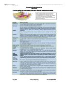
The differences in structure between the dendrites of a motor neuron and the sensory receptor of a somatosensory neuron are intimately related to their respective functions. In a motor neuron, the function of the dendrites is to pick up the neurotransmitters released from the presynaptic membrane of the preceding axon, and this is shown in their structure, a distributed field of complements to the synaptic knobs that they interface with. In a sensory neuron there are many different possible sensory receptors but they work under the same principle, a sensation detected in the receptive field is converted into an electrical signal. The most well-studied sensory receptor is the Pacinian corpuscle[1], shown in the sensory neuron diagram above. This elongated sphere of
wrapped connective tissue initiates a change in ion concentrations by the opening of channels when it is deformed by pressure. If the deformation is great enough and so enough of a change is made to the charge in the cell then an action potential will fire. Somatosensory neurons are unique in that they are adaptive for example, in the Pacinian corpuscle, a sustained pressure results in a change in the shape of the corpuscle as the layers of tissue slide over each other which closes the ion channels and so stops the triggering of action potentials after the initial signal.
As both types of cells are involved in the transmission of information over long distances, maximal use is made of the length of their axons and dendrites. It is for this reason that the synapses involving these neurons are either axodendritic, or they use the full length of the communicating neurite connected to the somatosensory neuron or motor neuron.
Both somatosensory neurons and motor neurons transmit their signal to its destination in a similar way, though there is an adaptation of the synapse at the end of the motor neuron which merits it a different name, a “neuromuscular junction” where the presynaptic terminals transmit a chemical signal to the motor end-plate which then effects the contraction. In a somatosensory neuron, the signal from the sensory receptor is conveyed along the cell, past the soma to the synaptic boutons at the axon terminal. Here, the charge triggers the release of neurotransmitter chemicals into the synaptic cleft where they diffuse over to be detected at the postsynaptic membrane of the dendrites on the other side. At a neuromuscular junction, neurotransmitters are released in the same way but they are picked up by receptors in the junctional folds of the motor end plate, with which the active zones are aligned.
2
Structural and Functional Differences Between Somatic Motor and Sensory Neurones Alex Baldwin
A similarity between somatosensory and motor neurons, noticeable under Golgi staining where the shape of the entire cell is visible, is that they both posses long neurites at each end, this is related to them both needing to send signals over long distances. Another similarity, visible under the SEM is that both types of cells contain many mitochondria. These mitochondria are necessary, in part, for the process of axoplasmic transport, which is the movement of chemicals along the neurites
between the soma and terminal. When moving materials from the soma to the terminal this process is known as anterograde transport and is required to move enzymes and neurotransmitters to the ends of the axons.
There is a biochemical difference between motor neurons and somatic sensory neurons. Motor neurons are cholinergic whilst somatic sensory neurons use glutamate as their neurotransmitter. In motor neurons, the enzyme choline acetyltransferase is synthesized in the soma and then moved by anterograde transport to the terminal where it plays a part in the formation of acetylcholine from acetyl coA and choline. Glutamate, however, is an amino acid and so is abundant throughout the body.
The transmission of the signal along the axon, though operating using the same principles in all neurons, has its differences between both somatosensory and motor neurons and within the group of different types of somatosensory neurons. The speed of the signal travelling along a neuron is dependant upon the speed of the transmission at the synapses at either end and the actual velocity within the neurites which can be encouraged by increasing their thickness and myelination. Myelination is the action that glial cells known as Schwann cells have on the axon, wrapping it in
an insulative barrier that prevents charge leaking out of the axon. Both alpha motor neurons and alpha somatosensory neurons (those used for proprioception) have thick, heavily myelinated axons in order to give their signals the rapid transmission speed that they need, which is approximately
120m/s at its maximum for motor neurons and 119m/s for proprioceptive neurons. Next in order of speed are those neurons with beta afferent axons, these are used for the communication of touch sensations and transmit signals at around 76m/s. Gamma motor neurons and somatosensory neurons that detect temperature and “fast pain” are next, with thinner, less myelinated axons resulting in a slower signal velocity, they have maximum speeds of 36m/s. Finally, “slow pain” is sent via c axons. These are thin, unmyelinated and by far the slowest neurons with a speed of around 0.6m/s.
Both sensory neurons and motor neurons come in a variety of different types to reflect their varied functions. Motor neurons are either alpha motor neurons, which are triggers for the contraction of muscles to move or manipulate the body and gamma motor neurons which are used to tense and shorten the specialised muscle fibres known as muscle spindles which are used to send sensory information about the load on a muscle at a specific time. If the gamma neurons were not there to tense the muscle spindles they would not stay sensitive when the muscle shortened which would mean that proprioceptive information on muscle stretch would not be sent.
There are many more different types of somatosensory neurons than motor neurons. These include those specialised for touch, proprioception (including muscle stretch), temperature and pain detection. These groups are then further divided into different types of neuron. For example, the touch sensor group is populated by Pacinian corpuscles (shown in the sensory nerve diagram above), Ruffini's endings, Meissner's corpuscles, Merkel's disks and Krause end bulbs. These different sensors are distributed, though not evenly, throughout the skin around the body.
One example where gamma and alpha motor neurons, and a somatosensory neuron come together is
3
Structural and Functional Differences Between Somatic Motor and Sensory Neurones Alex Baldwin
in the patellar reflex. This is a monosynaptic reflex where a strike from a tendon hammer causes the lower leg to kick out. When sat down at rest, the gamma motor neurons have the muscle spindles in the knee taut and ready to receive sensory information about the load on the muscles in the knee. When the hammer hits the tendon below the patella it stretches the muscle spindles in the quadriceps which sends an action potential along an Ia axon to a synapse in the grey matter of the spinal cord. Here, it interfaces with an alpha motor neuron which innervates the quadriceps muscle, causing it to tense and raise the lower leg at the knee, completing the reflex arc.
In conclusion, the structure and function of somatic sensory neurons and motor neurons differs mainly in ways that can be directly related with their function. However, they are still similar enough to bring to mind the basic origins of the nervous system, from simpler forms in our ancestors to its primal roots in the charges over the membranes of the prokaryotes from which life has evolved. Differences in placement can be seen that reflect the respective needs of the neurons to communicate to and from different cells whilst differences in morphology and in the supportive glial cells can be seen which show what the cells need to perform their function.
4
Structural and Functional Differences Between Somatic Motor and Sensory Neurones Alex Baldwin
References
Motor and Sensory Neuron Diagrams (Figures 1 & 2) Anon. Seen: 13 November 2006
Neuron Speeds Anon. Seen: 14 November 2006
Mark Bear, Barry Connors, Michael Paradiso. Neuroscience: Exploring the Brain
[1] – Mark Bear, Barry Connors, Michael Paradiso. Neuroscience: Exploring the Brain, p389
Motor Neuron Speeds
Granit, R. 1970 The Basis of Motor Control. London and New York: Academic Press. Cited By
Seen: 16 November 2006
5