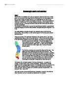
The skull is the complete skeleton forming a framework. It consists of a mosaic of many bones most of which are paired. These bones fit closely together to form a single rigid structure. It is basically a bone case around the brain, which provides holes for cranial nerves to exit and some vessels to enter the brain. Sensory organs are located in special compartments. For example: Eye - bony orbit
The main function of the skull therefore is protection, however, the skull has other functions such as to provide an attachment for muscles.
Protection: The cranial capsule encloses and protects the brain and important parts of the CNS. It protects by resisting deformation by strain put on it whether it be from the environment or internally such as food in the mouth.
There are five pairs of false ribs. The first three pairs attach to the sternum indirectly, and the last two pairs (the floating ribs) have no attachment to the sternum at all. There are seven pairs of true ribs, which attach directly to the sternum by the costal cartilages. The ribs help protect out vital organs such as our lungs and heart, it the chest its familiar shape. The ribs serve several important purposes. They protect the heart and lungs from injuries and shocks that might damage them. Ribs also protect parts of the stomach, spleen, and kidneys. The ribs help you to breathe. As you inhale, the muscles in between the ribs lift the rib cage up, allowing the lungs to expand. When you exhale, the rib cage moves down again, squeezing the air out of your lungs.
The appendicular skeleton includes the bones of the pectoral girdle, the pelvic girdle, the upper limbs and the lower limbs. The appendicular skeleton allows us to move around and do everything we need to do.
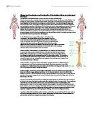
Just two bones make up the pectoral girdle. The clavicle (collarbone) and the scapula (shoulder blade). The clavicle is anterior and articulates with the acromion of the scapula and the sternal manubrium. The scapula is posterior and articulates with the head of the humerus.
right posterior right anterior
The pelvis is the bony structure located at the base of the spine. The pelvis incorporates the socket portion of the hip joint for each leg or hind leg. It forms the lower limb girdle of the skeleton.
The pelvis is symmetrical and each side is actually made up of three separate bones — the upper half, the middle and the bottom. These three bones fuse together with age. The pelvis is joined to the sacrum bone by ligaments and the hip bones nest in specially shaped sockets on each side. A female pelvis is wider and shallower than a male pelvis.
The pelvis protects the digestive and reproductive organs in the lower part of the body, and many large nerves and blood vessels pass through it to supply the legs. It is also an important load bearing part of the skeletal system.
Phalanges, metacarpals, radius, ulna, humerus, scapula, clavicle and carpals make up our upper limbs known as our arm, the function of the up limb is to help us with out every day activities. We use it to move things, to write and help do activities. We may use our arms to protect most parts of our body.
Femur, tibia, tarsal, phalanges and patella make up our lower limb which is known as our leg. We use our leg to walk.
Task 2
Joints are based upon their structure and function. The structural classification of joints breaks down into fibrous joints, cartilagenous joints, and synovial joints. The functional classification of joints breaks into the subdivisions of immovable, slightly movable or freely movable.
Fibrous joints connect bones without allowing any movement. The bones of our skull and pelvis are held together by fibrous joints. Cartilaginous joints are joints in which the bones are attached by cartilage. These joints allow for only a little movement, such as in the spine or ribs. Synovial joints allow for much more movement than cartilaginous joints. The hollow spaces between bones in synovial joints are filled with synovial fluid. This fluid helps lubricate and protect the bones. Examples can be your shoulder and your knee.
The ends of two bones which meet to form the joint are covered with articular cartilage. Its strength comes from tough fibers called collagen. The joint surface cartilage is well lubricated. The mature adult, articular cartilage has no blood vessels, no nerves, and no lymphatic channels. Its living cells are encouraged by joint fluid, called synovial fluid which is extremely good lubrication. In children, this cartilage does have some blood supply and can repair itself. In adults, the cartilage repairs with scar tissue which is mechanically lower.
Every synovial joint has an outer layer which looks like a sleeve. It is composed of strong, fibrous (collagen) tissue. Ligaments are a specialized part of this sleeve and account for the primary stability of the joint. The sleeve is oversized to allow for joint motion. It is encouraged by blood vessels which give it the ability to repair itself after injury. Joint lining produces synovial fluid, the prime lubricant for the joint and the nutritional source for joint surface cartilage and meniscus cartilage.
Synovial fluid removes impurities from the joint. It presents an enormous surface area. The lining acts as a two-way filter, allowing certain minerals, sugars, and proteins into the joints. Fortunately, most antibiotics pass through the synovial filter.
Ten percent of synovial joints have a washer like structure between bone ends called the meniscus. Its purpose is to absorb shock, to stabilize the joint, and to spread synovial fluid. It is made out of fibro-cartilage, which is a different tissue type from joint surface cartilage, but the meniscus also has no blood supply, no nerves, and no lymphatic channels. Biologically, it can't heal itself.
There are 6 types of synovial joints ball and socket, pivort, saddle, gliding hinge and candyloid. Synovial joints have bones covered at their articulating surfaces with articular cartilage (Hyaline cartilage).
- An example for a hinge joint will be your elbow. Hinge joints allows extension and flexion movements.
- An example for a ball and socket joint will be your hips and shoulders, a ball and socket joint allows for radial movement in almost any direction.
- An example for a pivort joint will be the neck and forearms. Pivot joints allow rotation around an axis. In the neck the occipital bone spins over the top of the axis. In the forearms the radius and ulna twist around each other.
- An example for a saddle joint will be the metacarpal bones in your hand they sit on top of each other allowing a back and forth and up and down movement
- An example for a gliding joint will be Metacarpals and midtarsal joints. It allows a gliding or plane joint bones slide past each other.
- An example for a candlyoid joint will be your wrist. Candyloid are similar to ball and socket joints. They allow the same type of movement but to a lesser magnitude.
Task 3