The leukemia will be harder to fight not only because of its new strength and resistance but the body is weaker from intense treatments. It can take a minimum of 5 weeks to start killing large numbers of leukemic cells and longer for the bone marrow to start growing back at a normal rate.2, 12 The overall cure rate in patients that have relapsed significantly lowers, varying between 80-10% in different cases. 13
Minimal Residual Disease & MRD Testing:
Even when in remission, everyone will still have a small number of leukemic cells in their bone marrow. This is known as minimal residual disease (MRD). The number of leukemia cells remaining is often less than 1 in 10,000, undetectable under a microscope. The MRD test detects the level of residual disease, leukemia, left with the patient and determines their risk of relapse. This is possible as the MRD test is a 100x more sensitive than a microscope.
If a patient relapses, the doctor explores the best option for the patient looking at higher doses of treatment as well as transplants.
By testing and recording levels of MRD within the blood, doctors are able to monitor each patient’s risk of relapse and tailor the treatment to suit them, giving intense treatment to those at high risk, saving those who aren’t the avoidable side effects.5
There are three main types MRD testing
DNA based tests: This aims to detect the Leukemic specific DNA sequence. The polymerase chain reaction is a technique of amplifying single or a few sections of a particular DNA section, a highly sensitive technique.
Specific markers are used for DNA based testing, these are chromosomal translocation for example t(14;18) involving BCL.
Chromosomal translocation is a chromosome abnormality caused by the rearrangement of different parts between chromosomes.
RNA based tests: Similar to DNA based tests but using RNA sequences. Reverse transcription of the RNA is used, followed by polymerase chain reaction.
For example, the t(9;22) BCR-ABL translocation may happen over a large length of the chromosome, making DNA based testing difficult. RNA is a much less stable target and requires careful handling and processing.
Patient specific testing: Uses immunoglobulin (IG) or T cell receptors (TCR) as a way of measuring levels of MRD in leukemia not containing a specific marker. The leukemic specific clone is enlarged and the specific region is arranged into a sequence. From this sequence PCR primers magnify the leukemic clone from the patient.
Both the DNA and RNA based tests require a pathologist to examine the bone marrow and decide which sequence to target. Once the target is found, a samples of blood or bone marrow is taken, nucleic acid is extracted, and the sample analyzed for the leukemic sequence.
MRD testing can be adapted for different leukemia’s and lymphoma, and research is currently being carried out to adapt the use of MRD testing in solid tumors such as testing the lymph nodes in breast cancer.13
There are currently a few controversies about MRD testing and whether it should be used for all patients, if there is a “safe” level of MRD and at what stages the test should be carried out. Through research is has been agreed that the test is carried out at diagnosis, 12-18 months into treatment and 24 months after the previous test. It has also been agreed that there will always be a tiny amount of residual left in the patient; however it would be best if as much MRD was eradicated as possible to prevent a high risk of relapse. 6
As MRD is £600 per test, it would be used as a routine test. In the long term, the cost of MRD testing for the NHS will be significantly lower than treating for relapse.4
Proof through Studies:
I have chosen three studies that prove the use of MRD testing, will benefit ALL patients by using MRD testing for both prognostic and preventative relapse treatment.
The first study I looked at was the “Importance of Minimal Residual Disease Testing, During the Second Year of Therapy for Children with Acute Lymphoblastic Leukaemia.” High levels of MRD after induction chemotherapy, shows the child’s level of resistance to chemotherapy and also the level of risk for relapse. It is so far unclear how helpful testing for MRD, in the second year of therapy and whether modified levels of treatment can change the result for patients with detectable MRD.
They looked at the level of MRD in the bone marrow samples of 85 children at 1, 12, and 24 months from initial diagnosis using clone-specific PCR primers designed to detect clonal antigen receptor gene rearrangements. These children were part of a multicenter, randomized clinical trial, which, in the second year of treatment, compared a 2-month reinduction -reintensification therapy followed by maintenance chemotherapy with standard maintenance chemotherapy alone.
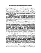
MRD was detected in 69% of patients at 1 month, 25% at 12 months, and 28% at 24 months from when first diagnosed. Through result analysis, high levels of MRD at 1 month, or the presence of any detectable MRD at 12 or 24 months from diagnosis, were highly predictive of relapse. Results showed, MRD testing at 1 and 24 months each had independent prognostic significance. Intensified therapy at 12 months from diagnosis did not improve prognosis in those patients who were MRD positive at 12 months diagnosis. The graph to the left helps show the results.
Clinical outcome in childhood ALL can be predicted with high accuracy by combining the results of MRD testing at 1 and 24 months from diagnosis. “Regular testing even after the patient achieves induced remission, will significantly lower the rate at which relapse is currently occurring.” This therefore proves that testing for MRD within the patient throughout treatment and remission will allow the doctor to adapt treatment for suitable levels for the patient and successfully predict the likeliness of relapse, and best way of prevention.
The second study looked at the clinical significance of MRD in childhood Acute Lymphoblastic Leukemia. This study investigated the significance of MRD in childhood acute lymphoblastic leukemia, looking closely at links and patterns that may arise between different patients.
Currently the connection and effect of residual disease after treatment of ALL is unclear. The Berlin–Frankfurt–Munster (BFM) treatment protocol was used, with minor modifications. Sequences of T-cell–receptor or immunoglobulin gene rearrangements (IG) were used as leukemic cell markers. Residual disease was measured using polymerase chain reaction assay.
A total of 246 children with ALL were enrolled in the study at the time of diagnosis, starting July 1993 and ending March 1996. Four patients were excluded because they did not reach remission, (fewer than 5% blasts in bone marrow smears). Sixteen patients were also excluded because less than three follow-up samples were obtained. 178 were monitored from start to finish with one or more clone-specific markers and an average follow-up period of 38 months.
The presence or absence and level of residual leukemia showed a strong positive correlation with the risk of relapse at each stage of monitoring. The PCR measurements recognized patients who were at high risk for relapse after the first stages of induction therapy. Analysis of the results showed that comparisons between immunophenotype, age, risk category (whether at standard or higher risk) and white blood cell count at diagnosis, the present level of residual disease were the single strongest prognostic factors for relapse.
Minimal residual leukemia found after initial induction treatment is a very successful method of gauging the level of relapse risk for childhood ALL patients. This means that the numbers of the relapse in children with ALL can be significantly lowered. “Detection of residual disease by PCR should be used to identify patients at risk for relapse and should be taken into account in considering alternative treatment. It is the single most powerful method in the prognosis of relapse.” The evidence collected shows that testing of MRD levels are the single most successful and reliable factor for the prognosis of the likelihood of relapse. The MRD test is also helpful in allowing doctors to tailor treatment doses to the patient, where previously everyone received the same dose. Sometimes doses may be too high, this way patients can be spared unwanted side effects.
Lastly I looked at a study investigating the prognostic role of MRD in Mature B-Cell Acute Lymphoblastic Leukaemia. The purpose of this study was to examine the impact of chromosomal translocation t(8;14) at diagnosis, and the response to treatment of MRD in B-ALL patients, on prognosis.
68 children affected by B-ALL were enrolled into the study for the Berlin-Frankfurt-Muenster LNH-97 clinical protocol. Bone marrow samples were taken from each patient and analyzed for the chromosomal translocation t(8;14)(q24;q32) by PCR. Samples were tested at diagnosis, after the first chemotherapy cycle, and after following cycles until each tested MRD negative.
47 patients (69%) of the studied were tested positive for t(8;14)(q24;q32). MRD response was shown in 39 patients, all of whom reached complete clinical remission and 31 in 39 patients became MRD negative after the first chemotherapy cycle. In patients that tested MRD positive after the first chemo cycle, the 3-year relapse-free survival was 38% compared to the 84% 3-year relapse free survival in those that tested MRD-negative. There was no difference in relapse free survival for children who reached a clinical complete remission after the first chemotherapy cycle (72%) compared to those who did not (79%), all shown in the results table to the right.
“Our study demonstrated that MRD carries a negative prognostic impact in B-ALL patients and suggests that a better risk-adapted therapy, possibly including the use of anti-CD20 monoclonal antibody, should be considered in selected patients.”
Although this study show that the MRD, currently only has a predictive value for success of treatment, it states in the results, that MRD testing significantly improves prognosis of relapse risk in ALL patients. It also showed how the MRD test may be adapted in the future for other types of leukemia or even cancers using PRC and translocation markers.
Source Evaluation:
I believe that all three studies that I have chosen to use are reliable and unbiased as they have been published in peer reviewed medical journals. Also all have received grants and funding from several well known medical institutions and charities.
Study one uses a sample size of 85, stating all methods and results clearly, highlighting the fact that everything was re-checked.
Study two enrolled 178 children, monitoring them constantly over a 3 year period and carried out follow-up checks up to 38 months later. They also use clear layouts and well explained graphs and tables to display their results.
Study three examined 68 children, the smallest sample size but reliable size. All information is proved in each stage, going into detail about how their results can adapted and carried out again to increases the success of prognosis of relapse, not only in ALL but may be other cancers.
Implications:
Economical: 10
On average, the NHS currently spends £110,000 on average for a full course of treatment leukemia per patient. This sum can significantly be reduced by using the MRD test, costing only £600 per test. Each patient, depending on their individual case, usually receives three tests over a period of 2 years. Using the MRD test will also reduce the amount spent and use of chemo and radiotherapy as patients may not need such strong doses for a long period of time before they reach remission, compared to the standard dose of treatment give to everyone.
By reducing costs for leukemic patients more money can be spent in other areas that need more attention like research for an translocation marker that can help adapt the MRD test for other cancers.
Social: 1, 14
MRD testing will reduce the amount of chemo and radiotherapy needed for some patients, sparing them the side affects, and will allow doctors to predict the likelihood of relapsing, which would save the lives of those who may not have been able to survive another attack from the leukaemia.
By inducing the remission stage of acute lymphoblastic leukaemia, it lengths the lives of the patients and allows them to return to normal life before the disease. Without reaching the remission stage, patients are required to spend a lot of time in hospital receiving treatment and recovering or at home resting. Receiving treatment takes a lot of energy and a long recovery period due to the side affects, which prevent the patient from socialising with friends and family.
Benefits & Risks:
MRD testing is a risk free procedure that only requires a sample of the patients’ blood, meaning that the test also has no side effects like the chemo and radiotherapy treatment.4, 5, 9, 11
Also, having the test allows doctors to tailor treatment to individual circumstances so not every patient has to go through such vigorous treatment.
The MRD test can also be adapted to detect minimal residual disease of different type of cancer. The MRD test for acute lymphoblastic leukaemia shows the B-lymphocytes and T-lymphocytes that have been affected, in breast cancer for example, they would be looking for a change in lymph nodes.
Treating children can be very difficult as their bodies are much more sensitive in comparison to adults. Therefore by using the same intensity of treatment on child that is used for adults may be successful but will in turn cause more damage to the child’s body than is necessary. Damaged areas that are of particular concern are the brain, heart, immune system, growth, toxicity and general development.4, 13
Alternatives:
There is not yet another alternative to relapse prevention, however the treatments that are currently being prescribed to treat ALL are sufficient enough to induce remission and the incidence rate of relapse is very low.
Relapse can be very serious as the leukemia comes back stronger, indifferent parts of the body and has usually become multi-drug resistant. If relapse does occur, or the patient is at high risk of relapse, as well as increased dosage of standard treatment; chemo with different drugs, radiotherapy, steroids, pain killers, immunotherapy etc, there are numerous other treatment possibilities. These are two options that are currently available. 1,2,3,14
- Prophylactic Cranial Radiotherapy (PCR) – when radiotherapy is given to a person’s head to prevent or delay the spread of a cancer to the brain. once the cancer reaches the brain and/or spinal cord, this is where it does the most damage.
When PCR is given, it is usually after chemotherapy, as the chemo drugs may not have been effective in treating any cells that may have spread to the brain. These cells may be too small to show up on a scan or to cause symptoms, so PCR is given as a preventative treatment to stop the leukemia causing problems in your brain later on. Sometimes chemotherapy is administered into the spinal fluid (intrathecal chemotherapy) as well as, or instead of, PCR.
Radiotherapy treats cancer by using high-energy rays to destroy the cancer cells with as little harm to normal cells as possible. PCR aims these high-energy x-rays at the head through a radiotherapy machine.
PCR is given as a series of short daily treatments in the radiotherapy department, using equipment similar to a large x-ray machine. Each treatment lasts for only a few minutes, the number of sessions vary depending upon each patient’s situation.
- Bone marrow or stem cell transplant – the replacement of healthy stem cells, allowing you body to produce normal, un-cancerous cells.
Bone marrow and stem cell transplants for ALL are intensive treatments. Intensive treatment is high dose chemotherapy, and sometimes total body radiotherapy. This treatment kills off all your bone marrow, containing the stem cells that make blood cells. The stem cells need to be replaced for the patient to survive treatment. You have the stem cells replaced by a drip of...
-
Someone else’s bone marrow or stem cells
- Your own bone marrow
- Your own stem cells, although this is very uncommon
(Illustrated in the diagram to the right)
The choice between a donor transplant and having your own bone marrow or stem cells depends on a number of different factors, including the type of leukemia and whether a member of family’s blood closely matches the patients.
Healthy marrow or stem cells collected and frozen until high dose chemo and radiotherapy are administered. They are then defrosted and given through a central line. The cells find their own way to the centre of your bones. They begin to make blood cells after a few days or weeks. Because of risk of infection, the patient must stay in isolation until the marrow or stem cells have started to make new blood cells again.
Bibliography:
E.g. Title - Date of last Access
Author
Source (Web Address/ Journal)
- Patient UK - 01/04/10
Multiple authors; certified by information standards.
(References Pui CH, Robison LL, Look AT, Crazzolara R, Bendall L, Belson M, Kingsley B, Holmes A)
- Cancer Research UK - 01/04/10
Author & References -
Page reviewed by: Dr P Mahendra, MD FRCP FRCPath
- National Cancer Institute – U.S National Institutes of Health - 01/04/10
Multiple authors from the department of health and human services and the national institute of health
- Leukemia & Lymphoma Research – Beating Blood Cancers - 01/04/10
Ken Campbell MSc, Clinical Information Officer
- Children with Leukemia - 01/04/0
Professor Mel. Greaves. Data from office of national statistics
-
The New England Journal of Medicine (Online) - 02/04/10
Multiple Authors: Hélène Cavé Cave, Ph.D., Jutte van der Werff ten Bosch, M.D., Stefan Suciu, M.S., Christine Guidal, M.S., Christine Waterkeyn, M.S., Jacques Otten, M.D., Marleen Bakkus, Ph.D., Kris Thielemans, M.D., Bernard Grandchamp, Ph.D., M.D., Etienne Vilmer, M.D., Brigitte Nelken, Martine Fournier, Patrick Boutard, Emmanuel Lebrun, Françoise Méchinaud, Richard Garand, Alain Robert, Nicole Dastugue, Emmanuel Plouvier, Evelyne Racadot, Alice Ferster, Jan Gyselinck, Odile Fenneteau, Michel Duval, Gabriel Solbu, Anne-Marie Manel, for The European Organization for Research and Treatment of Cancer–Childhood Leukemia Cooperative Grou
Supported by grants from the Association pour la Recherche sur le Cancer and the National Cancer Institute (5U10-CA11488-23 through 5U10-CA11488-28). Drs. van der Werff ten Bosch and Bakkus are the recipients of research awards from the Belgian National Fund for Scientific Research.
- Official Journal of the American Society of Clinical Oncology
Lara Mussolin, Marta Pillon, Valentino Conter, Matilde Piglione, Luca Lo Nigro, Paolo Pierani, Concetta Micalizzi, Salvatore Buffardi, Giuseppe Basso, Luigi Zanesco, Angelo Rosolen
Supported by Fondazione Città della Speranza, Associazione Italiana contro le Leucemie and by Camera di Commercio di Venezia.
Authors' disclosures of potential conflicts of interest and author contributions are found at the end of this article.
Volume: 25 Issue: 23 (20 Nov 2007) 5254-5261
-
Official Journal of the American Society of Clinical Oncology
Glenn M. Marshall, Michelle Haber, Edward Kwan, Ling Zhu, Daniella Ferrara, Chengyuan Xue, Michael J. Brisco, Pamela J. Sykes, Alexander Morley, Boyd Webster, Luciano Dalla Pozza, Keith Waters, Murray D. Norris
Supported by research grants from the National Health and Medical Research Council, Government Employees Medical Research Fund, and the New South Wales Cancer Council, Australia.
Volume: 21 Issue: 4 (Feb 2003) 704-709
- Kings College London – News Archive 09 - 02/04/10
Kate Moore, Public relations officer (Health Schools)
- BBC News – NHS Hidden Costs - 02/04/10
Government of National Statistics, The Treasury and the NHS
- Wikipedia – Minimal Residual Disease - 03/04/10
US National Library of Medicine
- Mac Millan Cancer Care & Support Charity - 03/04/10
- Mac Millan Cancer support bibliography
- Wikipedia – Acute Lymphoblastic Leukemia (ALL) - 03/04/10
-
CLIC Sargent – Caring for Children with Cancer - 03/04/10
Site terms and conditions page, source information
- Leukemia Research – Booklet - 30/03/10
Ken Campbell MSc, Clinical Information Officer
- Cancer Research UK – Leaflet - 28/03/10
Author & References -
Page reviewed by: Dr P Mahendra, MD FRCP FRCPath