One form of training used by athletes is called drop jumping and is classified as plyometrics (Fowler et al., 1994). The drop jump involves the athlete dropping from a predetermined height, and upon landing immediately performing a rebound jump. The training effect is generated by the rapid eccentric-concentric muscle contraction cycles (Boocock et al., 1990). Research has indicated that activities such as drop jumping apply significantly high joint forces about the ankle, knee and hip (Bobbert et al., 1987; Dufek and Bates, 1990), yet few studies have examined the implications of such training upon the spinal column. It is possible to reverse the effects of spinal shrinkage by adopting the ‘Fowler position’ where the trunk is supine, and the thighs are at a 40-degree angle with a bench or small chair supporting the lower legs. Studies such as Boocock et al. (1990) have used this method as it can quickly reverse the effects of spinal shrinkage, though may only be short lasting (Garbut et al., 1994). The magnitude of spinal shrinkage can be measured using precision stadiometry, this measure is considered highly accurate and has been used effectively by many investigators (Au et al., 2001; Fowler et al., 1994; Van Dieen et al., 1994). Furthermore, shrinkage in stature has been used as an index of spinal loading in both occupational (Cortlett, 1987) and sporting context (Reilly, 1988).
Widespread research into spinal shrinkage has been conducted over the past two decades that has resulted in the universal recognition of the shrinkage phenomenon (Boocock et al., 1988; Garbutt et al., 1994; Leatt et al., 1986). In 1987, Wilby et al. reported an average of 5.4mm of spinal shrinkage in the morning and 4.3mm in the evening over two circuits of eight exercises by females. The study not only recognises the phenomenon of spinal shrinkage, yet furthermore highlights the influence of exercise and circadian rhythms upon shrinkage. The implications of which can be readily explained in that the spine is at its most vulnerable in the morning (Reilly et al., 1984). Lying supine throughout the night relieves a proportion of the gravitational pull upon the spine, enabling the spinal column to return to its normal length. This results in the spinal column peaking in vulnerability early in the morning. Height is quickly lost during the first hour of waking and physical activities such as running and weight training have been shown to increase this normal rate of shrinkage (Leatt et al., 1986). In addition, Chambers et al. (2001) state that as people age their spines gradually shrink as the inter-vertebral discs lose moisture, slowly causing the discs to harden and therefore reducing mobility.
Research has indicated that spinal shrinkage occurs to a greater degree when load is dynamic not static (Tyrell et al., 1985). Therefore, activities such as plyometric training cause extenuated spinal shrinkage. Boocock et al. (1988) investigated the effect of dynamic loading on spinal shrinkage utilising a standing broad jump regimen and reported a shrinkage magnitude of 1.7mm. A more recent investigation by Boocock et al. (1990) investigated the stature changes following drop jumping. The protocol incorporated five sets of drop jumps from a height of 1m, rebounding over a hurdle 0.5m high. The regimen was of similar time duration and number of impact landings to the previous study, and yet reported a slightly greater mean shrinkage of 1.81mm over the two testing sessions. Thus indicating the drop jump regimen to greater influence shrinkage.
At present no investigation has sought to identify, the specific attributes of the drop jump regimen that greatest effect shrinkage. Therefore the study has been necessitated to investigate (1) the shrinkage induced by 2 separate drop jump regimens, and (2) to analyse the effect of drop jump acceleration upon spinal shrinkage.
Methodology
Participants
Six male subjects of mean (age 21+/-1.2yr, height 181+/- 6.3cm and weight 78 +/-8kg) took part in the study. All subjects took part in regular fitness training and had some previous experience of plyometric training. The subjects received an explanation of the protocol and provided informed consent before testing.
24 hours before each test the subjects were asked to eat and drink the same foods and were asked to refrain from exercise and also to go to bed at the same time.
Data collection
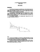
All subjects wore close fitting clothing and their own light indoor footwear. Prior to testing the subjects had joint centre markers placed on the shoulder, hip, knee, and ankle joints in the following positions; Shoulder- acromion process, Hip – lateral point of the greater trochanter (3cm superiorly, 1cm anteriorly); Knee – mid-point of the line through centres of the posterior convexities of the femoral condyles; Ankle – most distal palpable point of the lateral malleolus. Their stature was then measured using a recently calibrated (Holtain Ltd. Wales) stadiometer. The subjects then adopted the fowler position for fifteen minutes and performed A standardised warm-up was performed by each subject after which stature was again measured using the stadiometer. The subjects were then asked to perform a series of 5 drop jumps from 2 varying heights of 35 centimetres and bound up to a platform of 35 centimetres in height. The subjects were given the following verbal protocol to follow when performing the drop jump. Step off the box in a manner that is comfortable, land on the force reaction platform comfortably with both feet then with as little ground contact time as possible jump onto the second box. The boxes were 1.2 metres apart. This was repeated 5 times in quick succession with no rest period to simulate a typical set of depth jumps that are often part of any training programme. The subjects were asked to step off the box however they felt most comfortable onto a force reaction platform (AMTI manufacturers) sampling at 1000Hz interfaced with a PC running the AMLAB computer software package set to measure peak ground reaction force for each trial. The subjects had 7 days rest between trials in which they were instructed to perform no training and perform a minimum amount of physical activity.
As this particular exercise mainly involves body movements that occur predominately in the sagittal plane, a two-dimensional method of video capture was used as this was considered to be accurate for the present study. A Tripod mounted video camera (Panasonic AG-DP200) was used to collect video images of each subject performing the specified task left to right as specified and was set up following the BASES guidelines. The camera was set at a height of 78cm from the floor and positioned 4.81 metres from the centre of the force reaction platform and was parallel to the ground and perpendicular to the drop jump, this ensured the drop jump was performed centrally in the field of view. To ensure the drop jump was recorded accurately, all trials were recorded on VHS tape.
The video images were displayed and digitised using the DigeTEESer software programme. The joint centre markers were located on the screen and their centre was digitised.
Each point was digitised from when the toe left the box on the initial jump phase to when the toe was about to leave the force reaction platform on the jump up to the second box. DigiTEESer provided kinematic data for all the joints marked this data was then smoothed using a five point moving average. From this data peak hip velocity for each subject was calculated using the formula difference in hip vertical position/difference in time. A one way ANOVA was used to determine if any differences lie between block height and PGRF, PHV and stature change. Linear regression was used to determine if there was any significant relationship between the aforementioned factors.
Results
Table 1 indicates a block height comparison of subjects’ peak hip velocities (m/s-1) and peak ground reaction forces (N) reported in both drop jump regimens. Table 1 also shows the resultant stature change comparison between block heights reported immediately after the completion of each drop jump regimen.
Table 1: A drop jump height comparison of Peak Hip Velocity, Peak Ground Reaction Force and Stature Change
Figure 1: A mean and SD comparison of drop jump heights for peak hip velocities (m/s-1)
Figure 1 indicates the effect of which block height incurred upon the subjects’ peak hip velocities. A one-way analysis of variance revealed a significant main effect for block height on PHV reporting; F 1, 9 = 53.69, P < 0.01. Thus signifying that greater peak hip velocities were attained during the block 2 regimen when compared to the regimen of block 1.
A linear regression, Pearson’s test enabled an evaluation of the relationship between peak hip velocities (m/s-1) and block height among the subjects, and revealed a significant correlation, reporting; r = 0.933, P < 0.01. The result indicating a high correlation between peak hip velocity (m/s-1) and block height, thus suggesting increased block height, has the resultant effect of enhanced peak hip velocity.
Figure 2: A mean and SD comparison of drop jump heights for peak ground reaction forces (N)
Figure 2 indicates the effect of which block height incurred upon the subjects’ peak ground reaction forces (N). A one-way analysis of variance revealed no significant main effect (P < 0.05) for block height on PGRF, therefore indicating no significant difference between blocks 1 and 2 on peak ground reaction forces (N).
A linear regression, Pearson’s test was conducted between peak ground reaction force (N) and block height to assess the presence of a relationship between the two variables, and reported; r = 0.347, P > 0.05, thus revealing a non significant correlation between peak ground reaction force (N) and block height.
Figure 3: A mean and SD comparison of drop jump heights for stature changes (mm)
Figure 3 shows the effect of which block height incurred upon the subjects stature change (mm). A one-way analysis of variance revealed a significant main effect for block height on stature change (SC), reporting; F 1, 9 = 5.6, P < 0.05, therefore indicating that stature change was significantly greater following the block 2 regimen when compared to the regimen of block 1.
A linear regression, Pearson’s test enabled an evaluation of the relationship between stature change (mm) and block height, and revealed a significant correlation, reporting; r = 0.642, P < 0.05. Despite the significance level, the correlation reported between SC and block height was moderately low, therefore minimising the accuracy with which block height may predict stature change.
Figure 4: Two scatter graphs indicating the correlation between 1). Stature change (mm) and peak hip velocity (m/s-1). 2). Stature change (mm) and peak ground reaction force.
Figure 4 combines the stature change (SC) by 1). Peak hip velocity (PHV), and 2). Peak ground reaction force (PGRF) scatter graphs. A linear regression, Pearson’s test enabled an evaluation of the relationship between both SC by; PHV and PGRF, and revealed no significant correlation for either the SC by PHV, reporting; r = 0.436, P > 0.05, or the SC by PGRF, reporting; r = -0.230, P > 0.05. Therefore, the results indicate peak hip velocity and peak ground reaction force, are not significant predictors of stature change.
Discussion
The mean alterations in stature and hip velocity changed significantly with an increase in the height the drop jump was performed from after the spine was elongated using the fowler position. The present study did not find a significant difference in peak ground reaction force (PGRF) between the heights. Also found was block height was not a sufficient predictor to stature change using linear regression. The present study reinforced findings from other studies that drop jumping leads to spinal shrinkage. (Leatt et al. (1986, Mcnitt 1993, Dufek 1990) The mean loss of stature due to spinal shrinkage from the lower block was 2.2mm and the higher block lead to a mean change in stature of 5.1mm. The increase in hip velocity was due to the increase in block height, this lead to greater, although non significant, PGRF between the blocks and resulted in a significant change in stature due to the increased landing forces being absorbed by the intervertebral fluid contained between the vertrebral discs. The non significant difference in PGRF in the trials may possibly be due to the landing techniques adopted by the subjects. The method a subject uses to land after performing the drop jump may have a direct effect on the forces produced when landing (PGRF) and therefore will affect the amount of force travelling to the spine and contributing to spinal shrinkage. This is due to the subject attempting to dissipate the forces generated when landing throughout the body, and not focussing the forces on one area which can lead to injuries of the lower extremities. James et al.(2000) Found that subjects who showed symptoms of overuse injuries developed greater forces when performing drop jumps than subjects who had no signs of overuse injuries to the lower extremities. General increases in peak ground reaction forces, peak joint moments, and powers with increases in landing height and stiffness were also found by Zhang et al (2000). The researchers also found the knee joint extensors were consistent contributors to energy dissipation, a shift from ankle to hip strategy was observed as landing height increased and the contributions of lower extremity joints to energy dissipation during landings also increased supported by Kovacs et al (1999), thus further highlighting subjects strategies to dissipate forces on landing.
Overall the study found that when the spine is elongated, drop jumping leads to spinal shrinkage and loss of stature implications of these findings are as follows. Due to circadian rhythms the length of the spine varies throughout the day. as identified in previous research spinal shrinkage is a factor in lower back pain (garbutt et al.1994), when performing drop jumping, the shrinking of the spine may lead to lower back pain as the intervertebral fluid has already been forced from the discs and further loads could cause damage to the discs.. Therefore the drop jumping or indeed any activity that may lead to spinal shrinkage should be performed when the spine is elongated by adopting a suitable position such as the fowler position or when circadian variances lead to an increase in spine length e.g. the morning. This will aid in avoiding injury as when the spine is already shortened the forces generated by activities such as drop jumping will be absorbed by the spinal discs and not the intervertrebral fluid as this has already been forced out of the spine due to daily activity.
As identified the landing mechanics may play a role in dissipating the forces generated when drop jumping to other areas. Further research should be conducted into the affect landing mechanics have on spinal shrinkage and the ground reaction force generated when hitting the floor.Also an optimal height for drop jumping should be found in order to maximise the training effects of plyometric training such as drop jumping but minimise the effects of spinal shrinkage.
References
Adams, M.A. and Hutton, W.C. (1983). The effect of posture on the fluid content of lumbar intervertebral discs. Spine, 8, 665-71.
Bobbert, M.F., Huijing, P.A. and Van Ingen Schenau, G.J. (1987). Drop jumping 1: the influence of jumping technique on the biomechanics of jumping. Medicine and Science in Sports and Exercise, 19, 332-8.
Boocock, M.G., Garbutt, G., Linge, K., Reilly, T. and Troup, J.D.G. (1990). Changes in stature following drop jumping and post-exercise gravity inversion. Medicine and Science in Sports and Exercise, 22, 385-90.
Boocock, M.G., Garbutt, G., Reilly, T., Linge, K. and Troup, J.D.G. (1988). The effects of gravity inversion on exercise-induced spinal loading. Ergonomics, 31, 1631-7.
Caster, B.L. and Bates, B.T. (1995). The assessment of mechanical and neuromuscular response strategies during landing. Medicine and Science in Sports and Exercise, 27, 736–44.
Chambers, R., Hawksley, B., Smith, G. and Chambers, C. (2001). Back Pain Matters in Primary Care: Clinical Management of Back Pain in a Healthy and Safe Environment. Oxon: Radcliffe Medical Press.
Au, G., Cook, J. and McGill, S.M. (2001). Spinal shrinkage during repetitive controlled torsional, flexion and lateral bend motion exertions. Ergonomics, 44, 373-81.
Corlett, E.N., Eklund, J., Reilly, T. and Troup, J.D.G. (1987). Assessment of workload for measurement of stature. Applied Ergonomics, 18, 65-71.
Dufek, J.S. and Bates, B.T. (1990). The evaluation and prediction of impact forces during landings. Medicine and Science in Sports and Exercise, 22, 370–7.
Eklund, J.A. and Corlett, E.N. (1984). Shrinkage as a measure of the effect of load on the spine. Spine, 9, 189-94.
Foreman, T.K. and Troup, J.D.G. (1987). Diurnal variation in spinal loading and the effects on stature: a preliminary study of nursing activities. Clinical Biomechanics, 2, 48-54.
Foster, C.A., Maisel, R.H. and Meyerhoff, W.L. (1981). Head and neck trauma: initial evaluation, diagnosis and management. Minnesota Medicine, 64, 85-90.
Fowler, N.E., Lees, A. and Reilly, T. (1994). Spinal shrinkage in unloaded and loaded drop-jumping. Ergonomics, 37, 133-9.
Garbutt, G., Boocock, M.G., Reilly, T. and Troup, J.D.G. (1994). Physiological and spinal responses to circuit weight-training. Ergonomics, 37, 117-25.
James, C.R., Dufek, J.S. and Bates, B.T. (1999). Effects of injury proneness and task difficulty on joint kinetic variability. Medicine and Science in Sports and Exercise, 31, 708-16.
Koller, W., Funke, F. and Hartman, F. (1984). Biomechanical behaviour of human intervertebral discs subjected to long lasting axial loading. Biorheology, 21, 675-86.
Kovacs, I., Tihanyi, J., Devita, P., Racz, L., Barrier, J. and Hortobagyi, T. (2000). Foot placement modifies kinematics and kinetics during drop jumping. Medicine and Science in Sports and Exercise, 32, 1833-44.
Kramer, J., Kolditz, D. and Gowein, R. (1985). Water and electrolyte content of human intervertebral discs under variable load. Spine, 10, 69-71.
Leatt, P., Reilly, T. and Troup, J.D.G. (1986). Spinal loading during circuit weight-training and running. British Journal of Sports Medicine, 20, 119-24.
Markolf, K.L. (1972). Deformation of the thoracolumbar intervertebral joints in response to external loads. Journal of Bone and Joint Surgery, 54A, 511-33.
McNitt-Gray, J.L. (1993). Kinetics of the lower extremities during drop landings from three heights. Journal of Biomechanics, 26, 1037–46.
Porterfield, J.A. and DeRosa, C. (1998). Mechanical Low Back Pain: Perspectives in Functional Anatomy: Second Edition. Philadelphia: W.B. Saunders.
Reilly, D.T. (1988). Dynamic loading of normal joints. Rheumatic Disease Clinic of North America, 14, 497-502.
Reilly, T. Tyrell, A. and Troup, J.D.G. (1984). Circadian variation in human stature. Chronobiology International, 1, 121-6.
Troup, J.D.G. (1965). Relation of lumbar spine disorders to heavy manual work and lifting. Lancet, 17, 857-61.
Tyrell, A.R., Reilly, T. and Troup, J.D.G. (1985). Circadian variation in stature and the effects of spinal loading. Spine, 10, 161-4.
Van Dieen, J.H., Creemers, M., Draisma, I. and Toussaint, H.M. (1994). Repetitive lifting and spinal shrinkage, effects of age and lifting techniques. Clincal Biomechanics, 9, 61-3.
Van Dieen, J.H. and Toussaint, H.M. (1993). Spinal shrinkage as a parameter of functional load. Spine, 18, 1504-14.
Wilby, J., Linge, K., Reilly, T. and Troup, J.D.G. (1987). Spinal shrinkage in females: circadian variation and the effects of circuit weight training. Ergonomics, 30, 47-54.
Zhang, S.N., Bates, B.T. and Dufek, J.S. (2000). Contributions of lower extremity joints to energy dissipation during landings. Medicine and Science in Sports and Exercise, 32, 812-9.