Enzyme activity can be affected by various aspects like other chemicals, pH and temperature. They can be deformed by the heat and other chemicals change to change their active site so they will not be able to work properly and catalyse chemical reactions properly, this can lead to malnutrition in living organisms. pH can affect the enzymes by deforming the enzyme in such a way that the chemical properties of the enzyme are changed. Most of these deformations are irreversible and that means they can cause serious deformities to the mental and physical state of the living organism and sometimes leads to death. [8]
pH affects the secondary and tertiary structures of enzymes due to the way that the protein structure is organised. The reason that pH affects enzymes so greatly is due to the presence of basic (-NH3+) and acidic (-COO-) groups on the amino acids that make up the proteins. One polypeptide chain may contain hundreds of these groups. This means that any change in the pH of the solution that the protein is suspended in may cause the ionic charge to change; this is magnified by the number of the residual groups present in every protein so the effect may have a big impact on the shape of the active site. If the active site of an enzyme is changed in any way, the reaction that the enzyme catalyses ceases to occur due to the substrate not being able to fit into the active site. An enzyme-substrate cannot be achieved. [9]
The active site is the binding site on the quaternary structure of the globular protein. The recognition of the substrate is dependant on the chemical properties of the binding site along with the shape that it holds. Residual groups necessary for successful interaction with the substrates are found on the active site and help the enzyme bind itself to the substrate molecule. This is usually made up of 3-12 amino acids and only occupies around 5% of the total space on the protein. [8]
Theories regarding the binding of the enzyme and the substrate have been put forward, the most common of them being the ‘lock and key’ model theory and the ‘induced fit’ model. [8]
The lock and key theory suggests that the substrate locates the active site of an enzyme and attaches it self to the binding site. Once at the binding site, the substrate molecule is rearranged. The rearrangement of the substrate molecule causes the break down of the substrate; the products of this then leave the active site as they are no longer attracted to the residual groups of the enzyme. [8]
The second theory, the induced fit model, explains that the substrate molecules induce a change in the quaternary structure of the enzyme when it combines with the enzyme. This then forces a chemical change in both enzyme and substrate which then rearranged the molecules. [8]
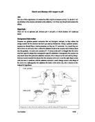
[12]
This diagram (above) shows how enzymes break down large insoluble molecules into smaller molecules where substrate is another name for the chemical that is going to be broken down, enzyme-substrate complex is another name for the partial molecule formed when the substrate and enzyme momentarily fuse. Products are what the substrate is broken down into.
What is Diastase?
Diastases are a family of enzymes that were discovered in 1833 by Anselme Payen. The name diastase is the generic name given to any enzyme in the amylase group; it digests amylase by hydrolysing the α-1,4-glycocidic bonds in the polysaccharide amylase. This breaks the starch molecules down into the smaller, more soluble maltose molecules. Diastases are active between pH 4.0 and pH 8.0; specific boundaries are varied by the enzyme. The different diastases are Alpha-Amylase, Beta-Amylase, Fungal Amylase, Glucoamylase and Malt Diastase. [2]
What is pH?
The pH scale measures the concentration of aqueous H+ ions (protons). This controls the acidity of solutions. Thomas Lowry and Johannes Brønsted independently came up with a definition for acids and bases in 1923. The Lowry-Brønsted definition for acids and bases is: “An acid is a substance that can donate a proton (an H+ ion) and a base is a substance that can accept a proton”. [10]
There is a conventional accepted definition for pH and that is “-log10 [H+ (aq)]”. The square brackets in this definition are there to show the concentration of H+ ions in mol dm-3. The pH scale depicts the concentration of ions present, as the number gets smaller, the concentration of H+ ions increases. A jump of 1, like from pH5 to pH4 for example, represents a tenfold increase in the concentration of H+ ions. [11]
What is a Buffer Solution?
A buffer solution is one that can resist changes to the concentration of the OH- ions and the H+ ions within the buffered solution. Buffer solutions usually withstand changes in pH when moderate amounts of acid or base are mixed within them. [11]
The pH in a buffer solution can be calculated using the Henderson Equation.
The Henderson Equation is:
Ka = [H+ (aq)] eqm [A- (aq)]
[HA (aq)] eqm
[11]
Apparatus and Glassware List
- 10 x Spotting Tile
- 2 x Spatulas
- 2 x Glass Rod
- Weighing scale accurate to 2 d.p.
- 4 x 200ml Beakers
- 2 x Funnel
- 5 x 250ml Volumetric Flask
- 1 x 500ml Volumetric Flask
- Safety Goggles
- Hot Plate
- Starch Powder
- Diastase Powder
- Wash Bottles containing Distilled Water
- 15 x Dropping pipette
- 6 x 25ml Pipette
- Stop Watch
- Thermometer
Making My Solutions
I will be making percentage solutions, 2.5% diastase solution and 1% buffered starch solutions. My solutions are going to be a set concentration and volume, which I will make uniquely for every pH buffer solution that I am going to make. I will then mix my buffered starch solution with my diastase solution and use a pipette dropper to drop drops of my solution into the spotting tiles already containing 2 drops of Iodine solution to determine the amount of digestion the enzyme has done to the starch.
Making pH Buffered Starch Solutions
- Rinse all your glassware and funnels with distilled water before using them.
- Weigh out 2.5g of starch solution in a beaker using the weighing by difference method.
- Dissolve your starch using buffer solution, making sure the starch is well dissolved.
- Heat the contents of the beaker at 70°C.
- Pour all the contents of the beaker into a 250ml volumetric flask via a funnel, rinse the beaker and rod and pour the solutions into the volumetric flask, then make up the solution to 250ml.
- Stopper and inverse the volumetric flask from 10-15 times making sure the solution has been thoroughly mixed.
- Repeat this procedure for all the different pH’s that you require taking care that no contamination of solution occurs.
NB: Allow the hot solution to cool before adding it to the diastase solution to prevent the heat from denaturing the diastase enzyme, measure the temperature with a thermometer
Making the Diastase Solution
- Weigh out 12.5g of diastase powder in a beaker using the weighing by difference method.
- Dissolved the diastase solution with distilled water.
- Pour the contents of the beaker into a volumetric flask, rinse all glassware, and pour the washings into the volumetric flask rinsing the funnel as well. Make the solution up to 500ml.
- Inverse the flask from 10-15 times to make sure it is thoroughly mixed.
- Keeping this solution in a refrigerator will not affect the enzyme so it should be possible to keep this solution for 48 hours and use it with other starch solutions. Be sure to bring the solution back to room temperature before using it as this change in temperature affects the pH of the starch solution.
Risk and Hazard Assessment
Starch:
Starch powder becomes very flammable if mixed with air and ignited, stay clear of any naked flames. You must always wear protective goggles whilst handling chemicals.
Diastase:
All enzymes are irritants. Do not inhale the dust or powder used, wear goggles and gloves when handling the solution. Appropriate protection must be worn; this includes safety goggles, loves and a laboratory coat. A mask can be worn if you are in doubt or if you are using large amounts of enzyme powder. Large quantities of enzyme powder are more likely to cause dust to be given off if disturbed in the slightest way.
Hot Plate:
Take care when using the hot plate. Keep your hands clear of the surface of the hot plate. There is a major risk of burning body parts that are exposed to the hotplate even if no contact is made.
Experimental Procedures:
- Place out two spotting tiles with two drops of Iodine solution in every dimple ready to test for starch in the mixture solution.
- Mix 25ml of the diastase and 25ml of the buffered starch solution into a conical flask.
- Swirl the mixture of solutions so that they are well mixed. Failing to mix the solutions properly will give you inaccurate results and ruin your investigation. This is because the starch will not have sufficient contact with the enzyme in some areas and will have a greater ratio or enzyme:starch ratio in other places. Start the experiment as quickly as possible to make sure you are getting as accurate of a result as possible.
- Drop two drops of the mixture into the dimples containing the Iodine solution every 30 seconds to test for starch in the mixture.
- Stop the experiment once the colour of the Iodine has stopped changing from orange-brown to blue-black. This indicates that all the starch has been digested.
- Repeat these procedures with all your different buffered solutions separately and record all your reading. You should repeat the experiment with each different buffer at least 3 times.
Analysis
Table of Results pH 3.45
Table of Results pH 4.35
Table of Results pH 6.27
Table of Results pH 7.17
NB: digestion did not occur at all for this pH.
Table of Results pH 9.22
NB: digestion did not occur at all for this pH.
The first thing that caught my attention was the results that I got for the tests I did using the pH 7.17 buffered solutions. I found that the enzyme that I used was inactive in pHs that are pH 7 and above. This tells me that the enzyme was denatured by neutral or basic conditions.
My results have thus shown me that the enzyme that I have been supplied with would have a range from pH 3 to pH 6, the specific enzyme can then be determined as Glucoamylase and that the range of this enzyme is pH 3 to pH 6. The optimum pH is somewhere in the boundary of pH 4.0 to pH 4.9.
Limitations to my experiments mean that I cannot guaranty this result although I can be certain which enzyme this is if I were to increase the number of trials that I did and if I use more accurate equipment and methods of measurement. I could also increase my experimental margin to the effect of pH on diastase in different temperatures.
Another thing that could have affected my results was the accuracy of my equipment. Values that I am aware of will always be different from the actual result. The 250ml and 500ml volumetric flasks I used to make my stock solutions had an error margin of ±0.05ml and the 25ml pipettes that I used to measure the solutions to mix has an error margin of ±0.06ml. One other error in measurement came from the weighing scale as I could only measure accurately to 2 decimal places.
After concluding my experimental procedures and analysing my results, I have decided that a more accurate range of pH’s is needed for better accuracy for my result. Decreasing the sample time from 30 seconds to 15 seconds will also help me achieve more accurate results.
Bibliography:
-
Hobby brouwen, published November 1999, picture is located midway down the webpage, , accessed 15th March 2008
-
enzyme India, published 2006, information located throughout the webpage in general, , accessed 14th March 2008
-
Wikipedia, published April 2008, information located at the top of the page, , accessed 2nd April 2008
-
Wikipedia, published April 2008, information located at the top of the page, , accessed 2nd April 2008
-
Wikipedia, published March 2008, information located throughout the page, , accessed 2nd April 2008
-
Wikipedia, published March 2008, information located throughout the page, , accessed 2nd April 2008
-
London South Bank University, published August 2007, , accessed 29th March 2008
-
Clegg C.J and Mackean D.G, (1994), Advanced Biology Principles & Applications, 3rd edition printed 1998
-
Howard R, (2002), Enzymes, Brian Ratcliff, Cambridge Advanced Sciences Biochemistry , 5th edition 2007
-
Ratcliff B and Eccles H, (2000), Acid, Bases and Buffers, Brian Ratcliff, Cambridge Advanced Sciences Chemistry2, 7th edition 2007
-
Lister T and Renshaw J, (2004) Acids, Bases and Buffers, Essential A2 Chemistry for OCR, 1st edition 2004
-
Gresham High School, Publishing date Unknown, , accessed 2nd April 2008