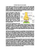
The function of an axon is that it Conducts nerve impulses
The function of a nerve terminal is that it Transmit information to neighbouring cells
- Synapse: A synapse is a junction (slight gap) between two connecting neurones. Impulses cross synapses by chemical means.
Types of Neurone
Sensory neurone, these carry sensory information from the external environment to the CNS
Sensory Neurone
The nerve impulses travel from left to right in this diagram of a sensory neurone. A stimulus causes the impulse to be produced by a sense organ. (Skin / ears / eyes / tongue / nose)
Dendrites and Synapses are both nerve endings at the ends of neurones. Dendrites are located at the ends that receive the nerve impulses (at the left of diagram above). Synapses are found at the transmitting ends of the neurone where the impulse is transferred to another neurone. Synapses use chemicals to transmit their electrical signal.
Motor neurone, this Carries information from the CNS to effectors organs
Motor Neurone
1. Nodes of Ranvier - constrictions of the myelin sheath which boost the passage of nerve impulses along the axon
2. Axon / Dendron - Axons carry the nerve impulses away from the cell body. Dendrons convey the electrical impulses towards the cell body.
3. Sheath - Layers of myelin wrap around the axon / dendron to protect it.
A motor neurone is connected to an effector and when an electrical nerve impulse is transmitted, the effector is stimulated into action. (Muscles / glands)
Inter-neurone (connector neurone, relay neurone, this is when small neurones making connections between neurones in the CNS
Relay Neurone
This neurone does exactly what its name suggests. Relay neurones are situated in the spinal cord. This, along with the brain, acts as the central nervous system.
Reflex actions are caused when a stimulus creates an electrical impulse that is relayed via the relay neurone straight to the effectors. The message never actually reaches the brain.
Glial Cells
Glial cells have a number of roles within the nervous system; they fill spaces, support neurones, enclose neurones and provide frameworks within nervous tissue. Glial cells are more abundant than neurones; there are ten times as many glial cells as neurones.
Glial Cells
- Astrocytes
- Epidermal Cells
- Microglia
- Oligodendrocytes
- Schwann cells
Schwann cells
- Cells which form the specialised myelin sheath of axons in the peripheral nervous system
- The plasma membrane may wrap itself around an axon up to 100 times, making the myelin sheath a white fatty material.
Summary of functions attributed to glial cells:
- electrical activity
- support nutrition of neurones
- secretion and storage of neurotransmitters
- proliferate to fill atrophied areas of the brain
- support for regeneration of damaged neurones
- mechanical support
- electrical insulation
- blood-brain barrier
- Control ionic composition of extra-cellular fluid.
Morphological nerve cell types
‘Networks’ of neurons
- neurons communicate across the synapse by using chemical messengers called neurotransmitters
- neurotransmitters may act to inhibit neurons or to excite neurons
- attachment of the neurotransmitters to pre-synaptic membrane receptors causes ion channels to open
•Information flow is usually in one direction (dendrites to synapses)
Gaps in the myelin sheath are formed between the bits of myelin provided by individual Schwann cells. These nodes of Ranvier are where the electrical signal is regenerated
Reflex action
- A reflex action is a very rapid automatic (no conscious control) response to a specific stimulus.
- Reflexes are involuntary actions that usually occur to aid the survival of an organism. A simple reflex is inborn and always results in the same response to stimulate.
- A reflex arc is the shortest pathway by which impulses travel from the receptor to the effectors in a reflex action. The simplest type is a monosynaptic reflex as it involves only a sensory and a motor neurone and hence one synapse.
- The parts of a reflex arc are:
1. Receptor (stimulated to generate impulse)
2. Sensory neurone, relay neurone in reflex centre (e.g. spinal cord) and motor neurone.
3. Effectors (muscle or gland stimulated to respond appropriately.
1. Spinal reflex actions: These are controlled by the spinal cord, i.e. their reflex centres are in the spinal cord. E.g. knee jerk and withdrawal of hand from hot or sharp objects.
2. Cranial reflex actions: These have their reflex centres in the brain (but the actions are NOT consciously controlled) .E.g. blink reflex, pupil reflex and tearing reflex.
- Simple reflex actions have a protective function. For example, the eye is protected by three simple reflexes:
1. Blink reflex (any object seen coming towards the eyes causes the eyelids to close).
2. Pupil reflex (strong light on retina makes pupils smaller to protect retina).
3. Tearing reflex (dust sensed by the conjunctiva causes an increase in tear flow to wash it away.
Features of the reflex include:
- They are very rapid, as they do not involve conscious thought.
- They are usually concerned internally with continuous and repetitive actions, such as breathing control of heart rate, and this frees the higher regions of the brain to deal with tasks involving complex thought.
The effect drug has on synapse:
A variety of drugs and poisons interfere with working of synapses. Some of these are agonists, e.g. they increase the activity of the synapse by inhibition acetylcholinestrerase or stimulating the action of the neurotransmitter. Others act as antagonists and so decrease the activity at the synapse; this is done by blocking the release transmitter or blocking the receptor sites. Many of these chemicals rely on the fact that shape plays an important role in the binding of transmitter and receptors, so molecules with similar shape interfere with the normal functioning of the synapse.
Nicotine is a similar shape to acetylcholinestrerase, so it can bind with ACh receptors and open sodium channels, so having a stimulating effect on the nervous system.
Beta-blockers have a similar shape to noradrenalin and compete with it for receptor sites so the ion channels remain closed and the impulse is not transmitted.
Prozac works as a serotonin re-uptake inhibitor. Serotonin is a transmitter normally active in the brain. Some forms of depression are caused by a reduced concentration of serotonin in the brain. Prozac competes with it for the re-absorption sites so more serotonin stays in the left. It also competes for the enzyme that normally breaks serotonin down.
Neurons transmit impulses as electrical signals. These signals travel very rapidly along their cell surface membrane. These signals are not a flow of electrons like an a electrical current but very brief changes in the distribution of charges cross the cell surface membrane, caused by the very rapid movement of sodium and potassium ions into and out of the axon. These movements are by diffusion and active transport.
Diffusion is when the ions move from high concentration to a low concentration active transport is when ions are pumped against the concentration gradient using energy from ATP.
Resting potential
In resting potential sodium/potassium pumps Na+ out and K+ into the axon, but the membrane is much more permeable to K+ so it tends to diffuse out again along the concentration gradient. Hence there are more positive ions outside than inside so the resting potential is -65 MV.
Action potential
In action potential sodium channels in he membrane open an N+ ions diffuse in rapidly along the concentration gradient. Now there are more positive ions inside than outside so the action potential is +40mv. The membrane is now said to be depolarised.
Return to resting potential
When returning to resting potential potassium channels in the membrane open and k+ ions diffuse out along the concentration gradient.
Bibliography: