purpose are called eluents. Polar eluents are not held strongly by the adsorbent and are preferable for
timely trials. Acetone, ethanol and methanol are the more polar eluents used in paper chromatography.
Less polar eluents include petroleum ether and cyclohexane. A problem that commonly arises in
chromatography is that the eluent is too strong for the mixture. If the solvent is too polar, it may move
the entire mixture with it. Trial and error is commonly used to determine the best eluent to use for a
specific mixture. Complete separation of the components of the mixture allows the retention factor (Rf)
to be calculated. The Rf value is the distance traveled by the substance divided by the distance traveled
by the solvent (Zubrick, 1992). However, running conditions greatly affect Rf values. Instead, a
reference compound should relate the positions of the experimental compound with others. Mobility of
individual pigments is affected by the degree of saturation of the tank with solvent, the type of paper
used, whether it is equilibrated with the solvent, and the amount of pigment applied (Goodwin, 1965).
For chlorophyll, up to eight colored spots can be observed. In order of decreasing Rf values they are
carotenes (two spots, orange), chlorophyll a (blue-green), the xanthophylls (four spots, yellow) and
chlorophyll b (green).
During storage chlorophyll encounters many elements which cause degradation. Chlorophyll a
is formed about five times faster than chlorophyll b. At room temperature in vitro chlorophylls will form
isomeric chlorophylls a and b, which are formed in vivo by heating. An in vitro sample will experience
isomerization in a few hours or rapidly if alkali is added at room temperature. Chlorophyll a and
chlorophyll b each have three isomers with magnesium bonded to three different pairs of pyrrole
nitrogen atoms. These isomers exist as tautomers rather than as resonant hybrids (Goodwin, 1965).
Also, the original chlorophyll sample may undergo oxidation and produce oxidized products.
The isomers and oxidized products account for the chlorophyll-like products that are separable from
chlorophyll on chromatographic analysis and they absorb at slightly different wavelengths. Oxygen
attack on the isocyclic carbon C-10 also breaks down the pigment. This process causes oxidation of
C-10 to hydroxy, then a breaking of the ring to form a variety of purpurins and chlorins. Further
oxidation of these derivatives occurs through complete scission of the isocyclic ring, then oxidation of
tetrapyrrole occurs (Goodwin, 1965). Other derivatives such as pheophytins a and b, pheophorbides
a and b, and pyroderivatives are caused by acidification, hydrolization and heat treatment, respectively
(Gross, 1991).
Reduction occurs not only by oxidation, but also by changes in temperature, pH and light.
Heating degrades the structure and instigates chemical reactions as well as further oxidation. The pH
changes during storage and processing leads to chlorophyll degradation. Chlorophyll stability varies as
a function of pH. A pH between 6 and 7 is critical. For spinach the critical pH is between 6.7 and 7
(Gross, 1991). Strong light bleaches chlorophyll solutions (Paech & Tracey, 1955). In vitro,
photobleaching causes chlorophyll breakdown where the chlorophyll solution is irreversibly bleached by
light in the presence of oxygen. Chlorophyll a and b have reaction rates on the same order. The order
of magnitude is larger than that of pheophytins and pheophorbides which are more stable. The
difference relates to the magnesium attached to the chlorophyll a and b excited triple state molecules.
Dim light studies during senescence show that the ratio of chlorophyll a to other chlorophylls decreases
while held in darkness. In this environment, direct photochemical degradation of the pigment occurs.
Spinach is a commonly used sample for determining the absorption spectrum of chlorophyll.
Chromatography can separate spinach into its chlorophylls a and b and the following carotenoids: ?-
carotene, lutein, violaxanthin, antheraxanthin, neoxanthin, zeaxanthin in order of increasing polarity
(Gross, 1991). For fresh spinach samples, the chlorophyll a to b ratio is 4.02 ? 0.79. With storage,
this ratio can be expected to change due to the faster degradation of chlorophyll a (Goodwin, 1965).
A single study has found okra to have a rather low chlorophyll a : chlorophyll b ratio of 1.208
(Gross, 1991).
1. Spinach leaves
2. Okra Pods
3. Distilled Water
4. 100% Acetone
5. Chloroform
6. 100% Ethanol
7. Whatman Chromatography Paper (2cm x 100m reel, 3mmChr)
8. Circular glass cuvettes
9. Square polystyrene cuvettes
10. 50ml polypropylene tubes (with caps)
11. Micropipette
12. 100 ml measuring cylinder
13. Aluminum foil
1. Milton Roy Spectronic 20D Spectrophotometer ( range: 400-700 nm, wavelength accuracy = ? 3
nm, absorbance accuracy = 0.5 percent transmittance)
2. Lab View (Interface designed by Al Giandomenico)
3. IEC -7000 Centrifuge
4. Absocol Refrigerator
5. Ultraviolet Lamp
The spinach leaves were cut into small pieces which were then ground with a mortar and pestle.
Okra pods were freed from seeds, cut into small pieces and similarly ground with a mortar and pestle.
The plant pulp thus obtained was then shaken with the solvent in a 50 ml polypropylene tube, whose
sides were wrapped with aluminum foil to minimize exposure to light. A volume of roughly 5 ml solvent
per gram of plant pulp was used. With the okra, it was necessary to leave the sample standing for at
least an hour to extract a substantial amount of chlorophyll. However, for spinach, shaking with the
solvent for 1-2 minutes would yield a relatively large amount of chlorophyll. The extract was then
centrifuged in the IEC- 7000 at 12?C at 3000 rpm for 15 minutes (a longer time of 20 minutes was
used for the okra extracts). The residue was discarded and the supernatant was transferred to another
50 ml tube (with the sides wrapped with aluminum foil) which was promptly shut to minimize
evaporation of solvent. With spinach extracts, 20 fold dilutions usually gave absorbance peaks within
the range of the spectrophotometer while no dilution was needed for the okra extracts. The cuvette
containing the sample was sealed using aluminum foil and adhesive tape and absorbance readings were
then taken over the 400-700 nm range with more data points being taken in the region of the peaks of
chlorophyll a and b. Four separate extractions of both spinach and okra were performed and the
resulting absorption spectra were recorded. The different spinach spectra were compared by
normalizing the peaks in the blue (430 nm) region and a similar normalization procedure was used for
the four okra extracts. One spinach extract and one okra extract were randomly chosen and the spectra
were compared using normalization.
To verify that acetone would be the most appropriate solvent for chlorophyll extraction, the
absorption spectra obtained with spinach extracts having water and acetone as solvents were recorded.
Equations (2) and (3) were available to determine the absolute concentration of chlorophyll a and b
from a 100% acetone extract using the absorbance readings obtained at 645 nm and 662 nm
(Lichtenthaler & Wellburn, 1983). However, these equations were valid only for 1 cm square cuvettes,
and only 1 cm circular glass cuvettes were available (acetone would dissolve the 1 cm square plastic
cuvettes). A comparison between absorbance readings from the two types of cuvettes was performed
using a spinach water extract to determine whether any conversion factor existed.
In order to verify whether the chlorophyll would degrade during storage in the refrigerator, a
refrigerated spinach extract obtained using 100% acetone was tested one week later using once again a
20-fold dilution and the two spectra were compared.
Paper chromatography was performed on one of the spinach extracts. A line was drawn
approximately 3 cm from the end of the chromatographic paper strip using a pencil. One drop of the
chlorophyll extract was applied at the center of the line using a micropipette. Approximately 5 ml of
eluent (both chloroform and ethanol were tested as eluents) was placed inside a 100 ml measuring
cylinder, and the end of the chromatographic paper strip was dipped inside the eluent, with the drop of
extract above the level of eluent. The cylinder was then sealed using aluminum foil and adhesive tape
and the eluent was allowed to move up the strip until the solvent front was near the top end of the paper
(Zubrick, 1992). The resulting chromatogram was viewed using an ultraviolet lamp. With optimum
separation, it is possible to see up to 8 bands showing the different chlorophylls, carotenes, and
xanthophylls.
Absorption Spectra of Chlorophyll from Spinach Leaves
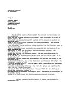
Figure 5 shows the absorption spectrum for one trial of spinach in 100% acetone. The ratio of
spinach leaf mass to acetone volume used was roughly 1 g : 5 mL (Appendix 1), and the extract was
diluted 20-fold for measurement taking.
Figure 5. Absorption Spectrum of Chlorophyll from Spinach Leaves
Figure 5 shows that the chlorophyll extracted from spinach has absorbance maxima in both the blue and
red regions of the visible spectrum. In the blue region chlorophyll absorbs maximally at (430 ? 3) nm,
while in the red region it absorbs maximally at (662 ? 3) nm. Also evident is a slight shoulder on the
peak in the blue region at a wavelength around 452 nm. The uncertainty in the wavelength setting for all
measurements with the Spectronic 20D is ? 3 nm, while the uncertainty in the absorbance
measurements comes from the Spectronic 20D’s uncertainty of 0.5 percent transmittance (Appendix 2).
Four separate extractions of chlorophyll from spinach leaves were performed, and one
spectrum was run for each of the extractions. The last three extractions came from the same batch of
spinach, while the first extraction was from a different batch of spinach. All four extracts were prepared
using roughly a 1 g : 5 mL ratio of spinach leaf mass to acetone volume and were diluted 20-fold for
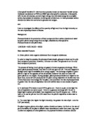
measurement taking. The four absorption spectra are plotted in Figure 6.
Figure 6. Comparison of Spectra from Different Spinach Extracts
The range in absorbance values at a given wavelength for the four spectra indicates that varying amounts
of chlorophyll were extracted. However, all four extracts had blue absorbance maxima of (430 ? 3) nm
and red absorbance maxima of (662 ? 3) nm (Table 1). Extract 1 was performed first, before
increasing the frequency of measurements, and thus it has fewer data points.
In order to accurately compare the spectra from separate extracts, it is necessary to normalize
the absorbance values. The spectra in Figure 6 were normalized using Extract 4, the one with the
largest absorbance values, as the basis. Normalization was done by multiplying the absorbance of each
data point of a given extract by the ratio of the peak blue absorbance of the basis to the peak blue
absorbance of the extract being normalized. The normalized spectra are shown in Figure 7 with error
bars omitted for visual clarity.
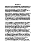
Figure 7. Comparison of Spinach Extract Spectra using Normalization
Figure 7 shows that the absorption spectra from the four different extracts of spinach agree very closely.
This indicates that the spinach data is very precise and that the acetone extraction procedure is
consistent in its removal of chlorophyll pigments from spinach leaves. Though the error bars are not
shown, the method for calculating the uncertainty of a normalized absorbance value is shown in
Appendix 2.
Absorption Spectra of Chlorophyll from Okra Pods
Figure 8 shows the absorption spectrum for one extraction of chlorophyll from okra pods using
100% acetone. The ratio of okra pod mass to acetone volume was 1 g : 5 mL, the same as for the
spinach leaves, however absorbance measurements were taken on the undiluted okra extract solution.
Figure 8. Absorption Spectrum of Chlorophyll from Okra Pods
Figure 8 shows that the chlorophyll pigments extracted from the okra pods for this trial absorb
maximally at (430 ? 3) nm and (662 ? 3) nm, just like the chlorophyll extracted from the spinach leaves.
Furthermore, the okra spectrum also has a slight shoulder in the vicinity of 450 nm, however it does not
appear as prominent as that of the spinach spectrum.
As with the spinach, four separate extractions of chlorophyll were performed for the okra pods.
The last three extractions were from the same batch of okra while the first extraction was from a
different batch. All four extracts were prepared using a roughly 1 g : 5 mL ratio of okra pod mass to
acetone volume, and the absorbance measurements were taken on the undiluted extract solutions. The
four absorption spectra that were obtained are shown in Figure 9.
Figure 9. Comparison of Spectra from Different Okra Extracts
The range of absorbance values at a given wavelength in Figure 9 shows that varying amounts of
chlorophyll were extracted from okra by the acetone. This is the same thing that happened with the
spinach. The first three extracts had blue absorbance maxima of (430 ? 3) nm while the fourth extract
had a maximum of (428 ? 3) nm. The red absorbance maximum ranged from (662 ? 3) nm to (665 ?
3) nm for the four extracts.
The spectra in Figure 9 were normalized using Extract 3 as the basis and are shown in Figure
10. The error bars are again not shown for the sake of visual clarity.
Figure 10. Comparison of Okra Extract Spectra using Normalization
Figure 10 shows that the absorption spectra from the four different okra extracts agree very closely, as
did the spinach extracts. This indicates that the okra data is very precise and that the acetone extraction
procedure is consistent in its removal of chlorophyll pigments from okra pods.
Comparison of Spectra from Spinach and Okra
Table 1. Comparison of Absorption Maxima
Blue Absorption
Maximum (nm)
Uncertainty in Blue
Absorption
Maximum (nm)
Red Absorption
Maximum (nm)
Uncertainty in Red
Absorption
Maximum (nm)
Spinach
1
430
3
662
3
2
430
3
662
3
3
430
3
662
3
4
430
3
662
3
Mean
430
3
662
3
Okra
1
430
3
663
3
2
430
3
662
3
3
430
3
665
3
4
428
3
664
3
Mean
430
4
664
4
Table 1 shows that there is no significant difference in either the blue absorption maximum or the red
absorption maximum for the spinach and okra chlorophyll extracts. The mean values for the blue
absorption maximum were (430 ? 3) nm for the spinach and (430 ? 4) nm for the okra. These are
identical to the blue absorption maximum of pure chlorophyll a. The mean values for the red
absorption maximum were (662 ? 3) nm for spinach and (664 ? 4) nm for okra, compared to the red
absorption maximum of 662 nm for pure chlorophyll a.
Since the four normalized spectra of spinach were virtually identical, and the four normalized
spectra of okra were virtually identical, as shown by Figures 7 and 10 respectively, any one of each can
be selected for comparing the spectra of the two plants. Figure 11 compares the second extracts of
spinach and okra. Both extracts were prepared with plant mass to acetone volume ratios of 1 g : 5 mL.
The spinach extract was diluted 20-fold for taking absorbance readings while the okra extract was not
diluted.
Figure 11. Spectral Comparison of Spinach and Okra
Figure 11 shows that at a given wavelength, the absorbance of the spinach extract is greater than the
absorbance of the okra extract even though the spinach extract had been diluted 20-fold and the okra
extract was not diluted. At the blue maximum of 430 nm, the spinach had an absorbance of 0.885
while that of the okra was 0.311. This indicates that either spinach has an intrinsically higher chlorophyll
content than okra does or else it is much easier for acetone to extract chlorophyll from spinach leaves
than from okra pods. The spinach leaves were thin and could be ground up easily, while the okra pods
had a tough, thick, fibrous skin, and could not easily be ground into small pieces.
The two spectra in Figure 11 were normalized using the spinach spectrum as the basis, and the
new curves are shown in Figure 12 below.
Figure 12. Spectral Comparison of Spinach and Okra using Normalization
Figure 12 shows that there is no significant difference between the normalized spectra of spinach and
okra. The points at 400 nm are significantly separated, however, this is at the lower bound of the usable
range of the particular spectrophotometer that was used. As one moves along the curves, at no points
does one curve deviate from the other. While it visually appears that the spinach spectrum has more of
a shoulder around 460 nm than does the okra spectrum, there is no statistical difference since the error
bars overlap. From the two spectra it can be inferred that the ratio of chlorophyll a : chlorophyll b is
very similar for the two plants. If one of the plants had a relatively larger amount of chlorophyll b than
the other, that plant’s spectrum would have more prominent peaks around 453 nm and 645 nm, the two
wavelengths at which chlorophyll b maximally absorbs.
In order to more accurately compare the relative amounts of the chlorophyll a and b pigments
in spinach and okra, a more quantitative means of comparison is required. Thus, equations (2) and (3)
were employed to determine the relative ratio of chlorophyll a to chlorophyll b using 100% acetone as
the solvent.
Ca = 11.75 A662 - 2.35 A645 (Equation 2)
Cb = 18.61 A645 - 3.96 A662 (Equation 3)
where Ca = amount of chlorophyll a in ?g /ml extract
Cb = amount of chlorophyll b in ?g /ml extract
These equations are valid for 1-cm square cuvettes, however, acetone degrades the square polystyrene
cuvettes that were available in the laboratory. Thus, round glass cuvettes had to be used for measuring
the absorbances of the extracts in acetone. However, for a given sample, round cuvettes give different
absorbance readings than square cuvettes since the round glass bends the light beam slightly. Since the
absorbance readings obtained in the round cuvettes will be different than those that would be obtained
using square cuvettes, equations (2) and (3) cannot be used to find absolute amounts of chlorophyll a
and chlorophyll b. In order to determine whether or not the equations can be employed to find the
ratio of chlorophyll a to chlorophyll b, an extraction from spinach using water as the solvent was
measured in both round and square cuvettes. This comparison will determine whether a fixed
conversion factor exists between each set of measurements.
Table 2. Comparison of Absorbance Readings in Round and Square Cuvettes
Wavelength
Absorbance
using round
1cm-cuvettes
Absorbance
using 1cm
square cuvettes
Ratio of
absorbances
Uncertainty in
Ratio
550
0.273
0.233
1.17
0.04
600
0.272
0.239
1.14
0.03
650
0.359
0.302
1.19
0.03
682
0.564
0.492
1.15
0.03
Table 2 indicates that for all four wavelengths tested, the absorbance in the round cuvettes was
significantly higher than the absorbance in the square cuvettes. Furthermore, the ratio of round to
square is not statistically different at the wavelengths measured as indicated by the overlap of their
uncertainty intervals (Appendix 2). Since equations (2) and (3) make use of absorbances at
wavelengths which differ from each other by only 17 nm (A662 and A645), assuming the same conversion
factor for both absorbances is reasonable. Both absorbance measurements taken for the equations will
be multiplied by the same factor to convert round absorbances to square absorbances. When Ca is
divided by Cb, the conversion factor can be factored out of both the numerator and the denominator.
Thus the ratio Ca : Cb obtained using the round cuvettes should be the same as that which would be
obtained using square cuvettes.
Using equations (2) and (3), the relative ratio of chlorophyll a : chlorophyll b was calculated
for each extract of spinach and okra, and the results are shown in Table 3 below.
Table 3. Ratio of Chlorophyll a : Chlorophyll b in Spinach and Okra.
Chlorophyll a : Chlorophyll b ratio
Uncertainty in ratio
Spinach
1
1.55
0.08
2
1.46
0.08
3
1.57
0.10
4
1.39
0.07
Mean
1.49
0.18
Literature Value
for fresh leaves
4.02
0.79
Okra
1
1.43
0.07
2
1.54
0.16
3
1.63
0.15
4
2.08
0.15
Mean
1.67
0.45
Literature Value
for fresh pods
1.208
only one study was
found
The uncertainties in the experimental mean values are the sums of the standard deviation and the
maximum uncertainty of one of the individual trials (Appendix 2). The literature value for spinach was
calculated from the results of three separate studies; Yamauchi et al. (1985), Khachik et al. (1986), and
Izaki et al. (1986) (Gross, 1991). The literature value for okra comes from a single study by Singh and
Dankhar (1980) (Gross, 1991). A paired t-Test between the experimental values of the ratios for
spinach and okra yielded a t-Stat value of -1.00, which is less than the t-Critical value of 2.35. This
indicates that there is no significant difference between the mean chlorophyll a: chlorophyll b ratios of
okra and spinach at the 95% confidence level. Table 3 shows that the experimental ratios are
significantly different from the literature values. The spinach extracts had a percent error of 67.1% while
the okra extracts had a percent error of 38.2%. However, it is very important to keep three points in
mind regarding the literature values. The literature values: (1) are for fresh samples, (2) are scarce, and
(3) show great variation. These points will be further discussed in the Discussion section.
Using equations (2) and (3) the absorbance values at 645 nm which are needed to obtain the
mean chlorophyll a: chlorophyll b ratios cited by the literature can be calculated. These values can
then be compared to the experimental absorbance values as shown in Table 4 below.
This allows verification of the changes needed in the absorbance values to cause significant changes in
the chlorophyll a: chlorophyll b ratio.
Table 4. Comparison of Experimental Absorbances with Expected Absorbances at
645 nm Given the Literature Chlorophyll a: Chlorophyll b Ratio
Absorbance at 645
nm Needed for Ratio
from Literature.
Experimental
Absorbance at 645
nm
% Deviation in
Absorbances
Spinach (literature
ratio = 4.02? 0.79)
1
0.138
0.221
-37.3
2
0.118
0.196
-39.6
3
0.085
0.135
-37.0
4
0.143
0.245
-41.5
Mean Deviation
-38.9
Okra (literature ratio
= 1.208)
1
0.222
0.200
10.9
2
0.080
0.069
15.8
3
0.099
0.083
19.5
4
0.075
0.070
7.5
Mean Deviation
13.4
Note the consistently larger absorbance values and the rather large mean deviation of - 38.9 % in the
case of spinach and the comparatively smaller mean deviation of 13.4% for okra.
Use of Paper Chromatography to Separate the Different
Pigments in Chlorophyll
Separation of the different pigments present in the leaf extract was attempted using ethanol and
chloroform as eluents. A good chromatogram would be valuable in confirming the presence of different
pigments in the leaf extract. However, no separation was observed when the chromatogram was
viewed with an ultraviolet lamp. This was due to the fact that both eluents were too polar such that they
would strongly bind to the different pigments and carry them all the way to the solvent front without
significant separation. Better results could be obtained using a less polar eluent such as petroleum ether.
Role of the Solvent in the Extraction Process
The choice of the solvent is a critical step in the extraction process. Chlorophylls being planar
organic compounds, are expected to be well solvated by acetone, which is a polar organic molecule.
Methyl alcohol or diethyl ether would also have made good solvents. In a very polar solvent such as
water, chlorophyll molecules associate to form dimers and polymers; resulting in marked differences
between the absorption spectrum as compared to the ones obtained using acetone. Figure 13
compares the absorption spectra of spinach extracts in water and in acetone.
Figure 13. Comparison of Absorption Spectrums in Water and Acetone
The spectrum in water has no real peak in the blue region and furthermore, the maximum in the red
region is shifted to the right to (678 ? 3) nm. This agrees well with the spectrum of chlorophyll
observed in a medium with a relatively large concentration of water molecules (Gurinovich & Pavlovich,
1971). Thus, for a better approximation of the absorption spectrum of chlorophyll which occurs in vivo,
using organic polar solvents is recommended since they solvate the chlorophyll molecules without
causing association and thus more closely replicate the conditions occurring in the leaves (Gurinovich et
al, 1971).
Storage
The chlorophyll content in plants is known to vary with time during storage with chlorophyll a
being degraded at a faster rate (Goodwin, 1965). To test whether the extracted chlorophyll would
remain stable over the span of a few days, a 100% acetone spinach extract was diluted using a dilution
factor of 20 and the absorption spectrum was measured on the same day. The extract was stored in the
refrigerator for one week and then tested one week later using the same dilution factor of 20.
Figure 14. Effects of Storage on Absorption Spectrum
Figure 14 shows that despite identical dilution factors, corresponding absorbance readings were
consistently higher after one week. One reason for this trend could be the rapid evaporation of the
acetone solvent (which has a relatively high vapor pressure) each time the tube containing the extract is
opened. This leaves behind a more concentrated extract of chlorophyll, thus accounting for the higher
absorbance readings. However, this was not an issue in subsequent tests with measurements taken
shortly after extractions, in sealed cuvettes to minimize evaporation of the acetone. Figure 15 compares
the two absorption spectra from Figure 14 using normalization.
Figure 15. Normalized Comparison of Storage Effects
Both graphs seem to agree at the blue and red peaks, which would suggest that the relative ratio of
chlorophyll a : chlorophyll b (which determines the shape of the graph, especially in the region of the
peaks) has shown little fluctuation over the one week of storage. However, higher absorbance readings
were obtained in the 500-625 nm region which raises the possibility that by-products which could be
formed from the breakdown of some of the pigments in the extract might be present after storage.
Discussion of Results
The absorption spectra of chlorophyll pigment extracts from both spinach and okra had
absorption maxima in two regions of the visible spectrum over the range of 400 to 700 nm. For the four
trials of spinach, the mean absorption maximum in the blue region was (430 ? 3) nm and the mean
absorption maximum in the red region was (662 ? 3) nm. The two mean absorption maxima for the
okra extracts were (430 ? 4) nm and (664 ? 4) nm. The small uncertainties indicate that there was an
excellent agreement between trials for a given plant and thus that the data were very precise. These
peaks are essentially the same as the absorption maxima of pure chlorophyll a, 430 nm and 662 nm.
Some of the absorption spectra had slight shoulders on the side of the blue peaks around 450 nm. This
is very close to the blue absorption maximum of pure chlorophyll b, 453 nm. Since the peaks of
chlorophyll a were more prominent, the spectra qualitatively indicate that the chlorophyll extracts from
both spinach and okra had relatively larger amounts of chlorophyll a than chlorophyll b.
The four extracts of spinach were all prepared similarly and diluted the same, yet for a given
wavelength, a range of absorbance values was observed for the four extracts. This same relationship
held true with the okra extracts. This indicates that for a given plant, varying amounts of chlorophyll
were extracted in the four trials. The amount of chlorophyll extracted depends on how finely the plant
sample is ground and on the length of time that the plant pulp spends in the acetone. The finer the plant
is ground, the greater the surface area over which the acetone can act. The longer the pulp is in the
acetone, the more time the acetone has to break the protein complex and remove the chlorophyll
pigments.
Though the various extracts for a given plant showed a range of absorbance values at a given
wavelength, when normalized, the spectra were statistically identical. At any given wavelength, the
uncertainty intervals on the four data points (since there were four extracts) overlapped. When plotted,
the points were basically on top of each other, but if they were not, their error bars overlapped. This
relationship held for the chlorophyll pigment extracts from both the spinach and the okra. This indicates
that for either plant, the acetone extraction procedure is consistent in its removal of chlorophyll
pigments.
The comparison of one spinach extract with one okra extract indicated that the two spectra
were not statistically different. It was expected that the two spectra would be significantly different since
spinach was expected to have a higher ratio of chlorophyll a : chlorophyll b than okra. The
experimental conclusion that the ratios were not different was supported by the results from employing
equations (2) and (3). Spinach had a mean chlorophyll a : chlorophyll b ratio of 1.49 ? 0.18 while
okra had a mean ratio of 1.67 ? 0.45. A paired t-test showed that the two means were not statistically
different at the 95% level of confidence. Thus, the spectra and equation results indicate that the ratios
are the same for spinach and okra.
The results from the equations were significantly different than the literature values. The
literature value for fresh spinach samples was 4.02 ? 0.79 while that for okra was 1.208 (only one
study found). Though the experimental values are very different than these values, it is important to
keep in mind three points regarding the literature values. First, the literature values are for fresh
samples. The research groups that performed these studies grew their own plants and tested the
samples immediately after collecting them from the plants. The plants at a grocery store were the only
ones available for testing. Thus the experiment was at the mercy of the grocer and the vegetable
wholesaler. Also the length of time that the plants had been stored was also unknown; a week or more
is not an unreasonable time period. Chlorophyll pigments degrade significantly during storage, thus
altering the ratios. Second, analytical literature data on the chlorophyll content of plants is scarce, and
most texts on plant pigments are quick to point this out. The literature value for spinach was calculated
from only three studies while that of okra came from only one study. Finally, the results of studies in the
literature show great variation. The standard deviation of the calculated literature value for spinach was
20%. Furthermore, many studies only gave absolute amounts of total chlorophylls rather than
breakdowns of chlorophyll a and chlorophyll b. Six such studies were found for spinach (Kaur and
Manjerkar (1975), two from Yamauchi et al. (1985), Khachik et al. (1986), Izaki et al. (1986), and
Ogura et al. (1987)) and they had a standard deviation of 20% (Gross 1991). For okra, two studies
were found (Singh and Dankhar (1980) and Gupta and Mukherjee (1981)) and they had a standard
deviation of 58%. Comparison of the experimental absorbances at 645 nm (used in equations (2) and
(3) to calculate the relative ratio of chlorophyll a : chlorophyll b) with the expected absorbances, as
shown in Table 3, revealed that the mean deviation in absorbances was -38.9 % for spinach. Such a
large deviation strongly suggests that the ratios experimentally obtained and those from the literature are
statistically different and probably not a result of the uncertainty in the spectrophotometer. The
experimental ratio of chlorophyll a : chlorophyll b (1.67) for okra was relatively closer to the literature
value and this trend is confirmed by the smaller mean deviation of 13.4% when the expected and
experimental absorbances at 645 nm are compared. However in the case of okra, it must be noted that
the absorbances in the red region were small (due to the relatively low concentration of chlorophyll)
extracted and thus, small changes in absorbance have a greater potential to cause relatively large shifts in
the ratio of pigments calculated. One way around this would be to use dilution factors (e.g. 10-fold
rather than 20-fold as previously used) which would maximize the peak in the red region for spinach, aid
resolution and thus minimize error in the calculation of the ratios. For okra, the problem lies in the length
of time required to extract the pigments. Allowing the okra pulp to stay in solvent overnight would
probably yield a higher concentration of extract.
Sources of error:
(1) The specific storage and growth conditions for the plants from which leaves (or pods) were taken
are completely unknown to us.
(2) Pheophytins a and b formed by the decomposition of chlorophyll a and b respectively are always
present in any extract (Paech &Tracey, 1955). Treatment of the chlorophyll with acid will speed up this
decomposition. In each case, the result is a shift of the blue peak to higher wavelengths and a shift in the
red peak to lower wavelengths.
(3) To accurately determine the chlorophyll ratio by spectrophotometry, the wavelength positions and
absorption coefficients used must be checked under the conditions of analysis. This is in order to check
any discrepancies in the coefficients found in the equation. This involves getting the spectrum for pure
known concentrations of chlorophyll a and b and then determining the absorption coefficients at the
wavelengths used in the equations.
(4) The derivation of equations (2) and (3) assumes that the absorbance of one of the chlorophyll
pigments would not influence the absorbance of the other. One way to verify this assumption would be
to mix known amounts of chlorophyll a and b and then determine whether the resultant spectrum is
indeed the mathematical sum of the absorbances of the individual pigments.
Modifications:
1. 100% extractions of the samples can be performed by adding more solvent to the residue of the first
extract and perform further extractions until no more chlorophyll is being removed from the sample.
Using square glass cuvettes, optimal use of the equations (2) and (3) can then be made to obtain the
absolute amounts of chlorophyll a and b as opposed to the relative amounts of the two pigments in
this case.
2. Similar equations as the ones shown for 100% acetone are available for other solvents such as
diethyl ether or methanol. These solvents can be used to perform extractions on a given sample and
the ratios obtained from the different equations can then be compared.
3. Equations (2) and (3) cannot be used to find the absolute concentrations of chlorophyll a and b
since circular cuvettes were used rather than the standard 1 cm square cuvettes used in the
derivation of (2) and (3). However the total chlorophyll content can be accurately determined by
measuring the magnesium content of the extract using an Atomic Absorption Spectrophotometer
(Tracey, 1955). This value can then be used with the ratio of chlorophyll a: chlorophyll b to find
the absolute concentration of each pigment. The total chlorophyll content in a leaf can also be
measured by recording the absorbance at a given wavelength where chlorophyll a and b are
known to have the same absorption coefficient (e.g. 602 nm for diethyl ether). This absorption
coefficient can be determined and absorbance readings can then be directly scaled to the total
chlorophyll content for a given extract.
4. To build a larger database of results two things can be done:
? The absorbance spectra of other plant samples could be measured to verify how far they differ from
okra and spinach.
? Further spectra of fresh samples of spinach and okra, grown under known conditions can be taken
in order to determine the effects of changes in temperature and light intensity on the total chlorophyll
content and the ratio of chlorophyll a: chlorophyll b.
5. Pure chlorophyll a and b can be mixed in the ratio determined for the given sample by equations
(2) and (3) and the resultant absorption spectrum obtained from this mixture can then be compared
to the one obtained for the sample. This procedure would serve as a check on the relevance of the
equation and its coefficients.
6. For smaller uncertainties in the relative ratio of chlorophyll a: chlorophyll b, dilutions, especially in
the case of spinach, can be performed such that the peak in the red region is optimized, even though
it is at the expense of having the peak in the blue region off-scale.
7. To show that the plant extract actually consists of a mixture of pigments, paper chromatography can
be performed with a less polar solvent such as petroleum ether (which was unavailable in The
Bioengineering Undergraduate Lab). With good separation up to eight spots corresponding to the
different pigments could be visible (Zubrick, 1992). Cutting the paper and eluting the separated
pigments would give extracts whose absorbances can be measured and compared to the known
spectra of the pure pigments in order to confirm identification.
Storage problems:
Chlorophyll a degrades faster than chlorophyll b to the corresponding pheophytin (Goodwin,
1965). The result is a lowering of the ratio of chlorophyll a: chlorophyll b with increasing length of
storage time of the plant. Furthermore, the total chlorophyll content of the plant will vary depending
upon environmental conditions such as amount of shading or season (Goodwin, 1965). From our
results, it did not appear that a storage time of a week would significantly alter the ratio of chlorophyll
a: chlorophyll b since the normalized plots for readings taken on the day of the extraction and one
week after the extraction were in close agreement with each other. However, evaporation of the solvent
appeared to be a problem since the extract became more concentrated. While this does not affect
determination of the ratio of pigments, it would significantly influence quantification of the total
chlorophyll content in the extract.
Absorption Spectrum in-vivo and in-vitro
While the absorption spectrum of chlorophyll gives an insight in its function during the process of
photosynthesis, it must be noted that it does differ from the spectrum in-vivo. For instance,
chlorophyll a has two peaks in-vivo in the red region, at 670 and 683 nm (compared to a broad 662
nm peak in acetone) while chlorophyll b absorbs at 650 nm in-vivo (compared to 643 nm in acetone).
These discrepancies are probably due to association between the individual chlorophyll molecules or the
complex formation with proteins (Goodwin, 1965). The presence of these different peaks for
chlorophyll a is related to the biochemistry of the photosynthetic process. P690 and P700 are two
specialized forms of the chlorophyll a pigments, with the numbers reflecting the peak absorbances in
vivo. Note that the absorbance in-vivo mentioned for chlorophyll a is a combination of all the various
specialized chlorophyll a molecules. P690 and P700 function as the primary pigments where the light
reaction of photosynthesis is actually driven. The other pigments, however, are accessory pigments
which absorb at higher energy and then eventually pass their energy to the primary pigments (Green,
Stout & Taylor, 1985). The fact that energy at either 690 nm or 700 nm (the upper end of the visible
spectrum) is needed to drive the reaction ensures a broader range of light available for photosynthesis.
Practical Application.
Research in the absorption spectrum of plants has been instrumental in elucidating the
biochemical pathways governing the photosynthetic process.
Figure 16. Comparison of Action Spectrum for Photosynthesis with the
Absorption Spectrum of Photosynthetic Pigments.
Figure 16, above, shows a comparison of the action spectrum for photosynthesis (which is a measure of
the rate of photosynthesis) with the absorption spectrum of photosynthetic pigments. It can be seen that
there is a direct correlation between absorption of light by the photosynthetic pigments and the rate of
photosynthesis. This effectively confirms that the pigments, in particular chlorophyll, are directly
involved in the photosynthetic process. Thus, with the rate of photosynthesis being dependent upon the
range of light available to plants, greenhouse owners can employ artificial lighting to ensure a continuous
supply of light to the plants from the appropriate region of the spectrum. For instance, budding,
flowering and fruiting in greenhouses are favored by a High Pressure Sodium Bulb which emits a
significant proportion of light from the red-orange-yellow region of the spectrum (Sierratech
International Homepage). On the other hand, a metal-halide (blue-green) spectrum seems to favor
healthy and strong vegetative growth (Sierratech International Homepage).
Campbell, N.A. Biology , 4th Edition, Menlo Park, CA: Benjamin/Cummings, 1996.
Goodwin, T.W. Chemistry and Biochemistry of Plant Pigments. New York: Academic Press, 1965.
Green, G.W and D.J. Stout. Biological Science. Cambridge, UK: Cambridge University Press, 1984.
Gross, Jeana. Pigments in Vegetables: Chlorophylls and Carotenoids. New York: Van
Nostrand Reinhold, 1991.
Gurinovich & Pavlovich. Spectroscopy of Chlorophyll and related compounds. Springfield, VA: U.S.
Atomic Energy Commission, 1971.
Lichtenthaler ,H.K., & Wellburn, A.R. Determination of total carotenoids and chlorophylls a and b
of leaf extracts in different solvents: Biochemical Society Transactions (11): 591-592
Paech K. and M.V. Tracey. Modern Methods of Plant Analysis. Berlin: Springer-
Verlag, 1955.
Sierratech International Homepage: http://www.grocell.com/docs/lighting.html
Wolf, Frederick T. Changes in Chlrophylls A and B in Autumn Leaves. American
Journal of Botany. 4: 714-717, March 24, 1956.
Zubrick, Jones W. The Organic Chem Lab Survival Manual, 3rd edition. New York:
John Wiley and Sons, 1991.
Appendix 1. Extract Preparation
Table A1-1. Amounts Used in Preparing Extracts
Extract
Mass of Plant Pulp Used
(g)
Volume of Acetone Used
(mL)
Spinach 1
10.169 ? 0.001
50 ? 2
2
6.231 ? 0.001
31 ? 1
3
6.810 ? 0.001
34 ? 1
4
6.072 ? 0.001
30 ? 1
Okra 1
7.621 ? 0.001
37 ? 1
2
7.338 ? 0.001
37 ? 1
3
7.546 ? 0.001
38 ? 1
4
7.523 ? 0.001
38 ? 1
Appendix 2. Error Calculations
UNCERTAINTY IN ABSORBANCE MEASUREMENTS
The Spectronic 20D manual stated that the inaccuracy of percent transmittance readings was less than
0.5%. Since the accuracy of the machine was given in terms of percent transmittance, it must be
converted to absorbance. Absorbance and percent transmittance are related as follows:
OR
Employing the differential approach to determine the maximum systematic uncertainty in the absorbance:
(Equation 4)
The uncertainty is calculated using ?%T = 0.5.
UNCERTAINTY IN NORMALIZED ABSORBANCE MEASUREMENTS
The normalization procedure employs the equation:
where AN is the normalized absorbance value
AB is the peak absorbance of the basis
AS is the peak absorbance of the spectrum being normalized
AO is the absorbance at the point being normalized
Employing the differential approach:
(Equation 5)
UNCERTAINTY IN RATIO OF CHLOROPHYLL a TO CHLOROPHYLL b
The ratio of Ca : Cb comes from equations (2) and (3):
Ca = 11.75 A662 - 2.35 A645 = C1 A662 - C2 A645
Cb = 18.61 A645 - 3.96 A662 = C3 A645 - C4 A662
Thus, the ratio is given by:
(Equation 6)
UNCERTAINTY IN RATIO OF ROUND: SQUARE CUVETTE ABSORBANCE
The ratio is given by:
where AR and AS are the absorbances in the round and square cuvettes
(Equation 7)
Appendix 3. Absorption Spectra
Figure A3-1. Spinach Extracts in 100% Acetone
Figure A3-2. Okra Extracts in 100% Acetone
Figure A3-3. Spinach Extract in Water
Group R1-1