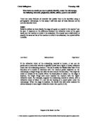
Diagram of the Becke Line Test
1 A thin section is a ground down slice of rock (to 0.03mm thick) which has been glued to a glass slide
Form and Cleavage
The form of a crystal is the shape it acquires as it grows. For example, crystals could be tabular, platy, scaly, rhombohedral, or rounded. Some crystals do not have a typical form. These crystals are described as irregular. Unfortunately, under the petrographic microscope, crystal form is not diagnostic. Crystal form needs to be considered along with another property for it to be diagnostic. An example of this property is cleavage.
Cleavage planes are lines of weakness along which the crystal would most likely break if hit with a hammer. The number of cleavage directions and the angles which they make with each other and the crystal faces can help to determine what a mineral is. Under the petrographic microscope, cleavage is seen as faint lines on the top of the crystal. A diagram is shown below to illustrate this.
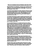
Extinction Angle
Extinction angle is the angle through which a crystal must be rotated for it to appear black (opaque) under crossed polars. When a crystal is placed in plane polarised light, it will rotate the light through an angle . This means that some light will be seen with the analyser in, when you would not expect anything to be seen if there was nothing there. The crystal will appear black under crossed polars when rotated through the angle . The angle is measured from the cleavage plane (or another linear feature). There will be an extinction position possible every . One point worth noting is that micas do not ever go completely black under crossed polars, instead there are tiny smudges of light going through. This is a diagnostic feature. When the extinction angle is , the extinction is described as straight. Other types of extinction that are possible are inclined, and symmetrical. Some minerals have no obvious extinction angle, whereas others are always in extinction. Minerals that are in permanent extinction are called isotropic.
Diagrams showing the measurement of extinction angle.
Colour
Another useful diagnostic property of minerals is colour. When looked at in a hand specimen, colour is not usually diagnostic. However, in thin section it can be. A lot of minerals are colourless in thin section, but some show quite distinct colours. For example, garnets usually show up pink in thin section and biotite usually appears brown or green.
Pleochroism
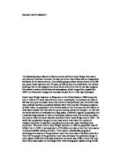
The colour of some minerals changes in polarised light depending on the angle at which they are orientated. This property is known as pleochroism. The colour varies with vibration direction due to the absorption of certain wavelengths of light. When the crystal’s slow vibration direction is parallel to the orientation of the plane polarised light there is a strong absorption and the colour is quite dark. Conversely, when the crystal’s fast vibration direction is parallel to the orientation of the plane polarised light, the absorption is much weaker resulting in a much lighter colour being observed. Taking biotite mica as an example, when aligned in the slow vibration direction the colour is dark brown but when aligned in the fast vibration direction it is light yellow. This is shown in a diagram below. When the vibration directions are at an angle to the polarised light orientation, the colour observed is somewhere in between the two extremes.
Pleochroism in biotite
Birefringence
One final diagnostic feature of minerals when under a microscope is birefringence. When a mineral is not in extinction, the plane polarised light from the polariser (E-W) is split into two directions when it enters the crystal. These two directions are perpendicular and they are along the two permitted vibration directions. Because these two components travel through the crystal at different speeds a phase difference develops between them, so they leave the crystal out of phase. This causes a characteristic colour, dependent on the phase difference, to be seen when the two rays are recombined by the analyser to vibrate N-S. The value of birefringence which is diagnostic for a mineral is the maximum value. The maximum value of birefringence can be determined by using a Michel-Levy colour chart. To use this, you look at the crystal with the analyser in, note the colours seen, and compare them to the Michel-Levy chart. The colour which appears furthest to the right on the chart corresponds to the largest phase difference. You then read from the scale to get a numerical value for birefringence. It is possible to have zero birefringence – this occurs when the vibration directions have equal speeds.
Twinning
Some minerals exhibit a quality called twinning. This is when a crystal has two or more adjacent parts where the crystal structure is differently orientated. The different parts of a twinned crystal are separated by a twin boundary. At the plane of contact, the crystal structure is similar for both components. Twins form when there are mistakes during crystal growth, for example mechanical deformation or a change in crystal structure as a result of cooling. Diagrams of some common forms of twinning are shown below.
Characteristics of plagioclase, biotite, olivine, quartz and calcite