We can identify that conscious perception lies in the brain by the observation that certain neuropathologies may impair it and that the introduction of certain drugs can alter an individual’s perception. This leads some people to think that the brain’s sensory apparatus functions to present the received information to an inner homunculus (see fig. 1); this concept is known as the Cartesian Theatre (Blackmore, 2005). This idea can be shown to be flawed on at least three levels. Firstly it does not address the actual function of perception; it is only moved to the “brain” of the homunculus. Secondly, this model implies that the brain works serially, processing information as a stream and presenting it to a central controller. Though this might seem plausible from our subjective experience the brain actually processes in parallel with no “pontifical cells”, as William James would put it, making all of the judgements and decisions. Thirdly, it can be demonstrated that the information that we act upon consciously in our brains is not a complete up-to-date representation of what we perceive with our sense organs. Some information can be shown to be absent or ignored whilst other areas may be confabulated or filled-in without our knowledge (Ramachandran, 1998).
The mechanism by which certain sensory inputs and mental processes are focused on has proved accessible to scientific research and is known as selective attention. Our ability to focus the brain on a particular task has been demonstrated to result in faster processing times and better results for that task (Womelsdorf et al, 2007). The question of how attention occurs has mainly been explored using the visual system. Experiments performed on binocular rivalry, where different images are shown to each eye so that they compete for attention, in monkeys have shown that changes in neuronal firing related to attention occur in the temporal cortex with the firing in the visual cortex remaining largely the same despite differences in what the subject is attending to (Sheinberg et al, 1997). Other experiments seem to show that selective attention relies upon the rhythmic firing of directly involved neurones with a higher degree of synchronization correlating with higher behavioural performance in tests performed on the visual cortex in monkeys, the rate of synchronization in this case being in the range of 40 to 100Hz (Womelsdorf et al, 2006). There is not yet any definite unified theory of how selective attention functions but experiments such as the above are key stepping stones towards further understanding.
A closer look can be taken at which areas of the brain contribute to conscious experience in the phenomenon known as blindsight. This is where some patients who are apparently blind or who have large blinded areas in their visual field can be demonstrated to be receiving some visual information without being consciously aware of it. The area most associated with conscious visual perception in humans is the striate cortex, also known as V1, with the left side of our visual perception mapping to the striate cortex of the right hemisphere and vice-versa. When damage occurs to the striate cortex patients report blindness in the part of their visual field mapping to the damaged area. This raised questions in neuroscientists who experimented on monkeys because when the striate cortex is damaged in the primate brain the subject can still perform in visual tests, though with some alterations in performance. The nature of the testing was hypothesised to be the cause of this difference; animals can’t communicate their conscious awareness of visual stimuli like humans can and this leads to any testing that is done on them relying on physical reactions to a particular stimulus. When humans with damage to their striate cortex are given similar tests to those performed on monkeys by forcing them to guess the nature of a stimulus that they’re exposed to they perform quite well, able to discriminate shapes, colour and movement. The differences in their performance are similar to those in monkeys with damaged striate cortex (Weiskrantz, 1997).
The usage of limited visual information without the associated qualia may have a particular significance due to the findings of Benjamin Libet. Libet demonstrated using electrical stimulation on the exposed cortex of his patients that conscious experience of being touched lagged behind the stimulation of somatosensory perception areas in the brain by about half a second (Blackmore, 2005). If there is a similar delay in our visual perception then some fast-track pathways for dealing with things we have to react very quickly to would be critical to our survival. Some would take this idea even further and hypothesise that our conscious perception of events in time is looser than we might think, illustrated by the example of the “cutaneous rabbit” where bunches of taps at the wrist, elbow and upper arm are felt as the evenly spaced movement, continuous in time from start to finish, of a “scurrying animal” (hence the name) up the arm of the subject (Rosenthal, 2005). This illusion demonstrates the brain retroactively filling-in points of conscious experience in time.
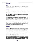
Experiments performed on epileptics who have had the tract of axons communicating between their cerebral hemispheres, called the corpus callosum, severed have provided some interesting information pertaining to how the two sides of the brain contribute to conscious experience. These patients appear to have no noticeable problems in their normal life but there are ways of exploiting the lack of the main route of communication between the two halves of their brain to perform experiments on them. Relying on the lateralisation of visual perception detailed above researchers could send information to each cerebral hemisphere separately. In one such experiment (see fig. 2) the word “key” was presented to the left visual field (to be processed by the right hemisphere) the patient could not see a word there as the areas for speech are centred in the left hemisphere in most people. However he could pick out the correct object from a collection in front of him with his left hand. When the word “ring” was presented to his right visual field however he recognised the word and could repeat it (Weiskrantz, 1997). In this way the two hemispheres could be seen as two separate consciousnesses. People believing that consciousness depends on language however may argue that only the left hemisphere in this case could be called conscious with the right hemisphere acting only subconsciously (Blackmore, 2005).
Anosognosia is a condition where the patient will be unaware of, or even deny, a sensory or motor impairment with the brain producing remarkable confabulations to protect this idea. An example can be found in a stroke patient who had a paralysis of the left side of his body. The patient claimed he was able to move his left hand and when he was presented with the lifeless article by the experimenter he reasoned that it was one of the experimenter’s three hands to go with the three arms he believed the experimenter to have (Weiskrantz, 1997). This denial can be averted temporarily however by injecting cold water into the ear of the patient. This can cause the patient to acknowledge their illness and even recognize that they have been paralyzed for some time but after the effect has worn off the patient will deny ever having admitted anything. The method by which this works is not yet known but is theorized to involve arousing areas of the brain that can recognize the problem with the affected part of the body (Ramachandran, 1998). What this does show however is that anosognosia has a definite neurological basis and is not a result of any higher level “repression”.
Another condition with implications for consciousness called unilateral neglect sometimes occurs after a stroke or lesion affecting the right parietal lobe. In these cases the patients will not only fail to consciously perceive visual information presented in the left side of a visual stimulus (though not reporting any blind areas) but also fail to produce information related to that left side. An example of such a case was in an experiment by Edoardo Bisiach where he asked them to describe an area that they knew from the perspective of entering it from one end. The patients neglected to mention any information pertaining to the left side. However when the patients were asked to imagine entering from the other end they spoke about all of the buildings that they did not before but this time neglected the other side of the area that would now be the left hand side (Blackmore, 2005). Other experiments have shown that there is a subconscious awareness of the neglected information. Stimuli presented to the neglected field facilitated a faster response when the stimulus was then presented to the side that the patient was aware of (Weiskrantz, 1997).
The scientific analysis of consciousness is still in its infancy but the progress in understanding that has already been made is encouraging. Further evidence from neuropathological cases may yet shed more light on our understanding of consciousness. A condition known as Akinetic Mutism results from damage to the intralaminar nucleus or anterior cingulate gyrus. In these cases the patient is awake and absorbing information and may even move if prompted by pain but will undertake no actions of their own volition. Interestingly, having recovered they say that they felt absent of thought during the experience which may be of great significance but we do not yet know the details of how their consciousness is altered during the state (Ramachandran, 1998). Further questions can be asked of areas away from the biology of living organisms. Could a non-living assembly that processes information such as a computer or network of computers become conscious? And if not what is it about the cells of our nervous system that makes them so special? In any case, there is obviously something particularly special about our drive to understand consciousness. In doing so we are effectively applying the most complicated tool we know towards studying itself, though whether this recursion will preclude any meaningful answers being found is uncertain.
References
Banks, W.P. (1995)
Evidence for Consciousness
Consciousness and Cognition vol. 4 pages 270-272
Blackmore, S. (2005)
Consciousness: A Very Short Introduction
Oxford University Press, Oxford
Blackmore, S. (undated)
Consciousness: An Introduction. Illustrations
http://www.susanblackmore.co.uk/Books/Consciousness/illustrations.htm accessed 05/03/2008
Churchland, P. M. (1988)
Matter and Consciousness
The MIT Press, Cambridge, Massachusetts
Cole, M. (2007)
History’s Top Brain Computation Insights: Day 15
http://www.neurevolution.net/ accessed 05/03/2008
Koch, C. and Greenfield, S. (2007)
How Does Consciousness Happen?
Scientific American vol. 297 issue 4 pages 76-83
Miller, S. M. (2007)
On the correlation/constitution distinction problem (and other hard problems) in the scientific study of consciousness
Acta Neuropsychiatrica vol. 19 pages 159-176
Qiu, J. (2007)
Probing Islands of Consciousness in the Damaged Brain
Lancet Neurology vol. 6 pages 946-947
Ramachandran, V. S. (1998)
Phantoms in the Brain
Harper Perennial, London
Rosenthal, D (2005)
Consciousness and Mind
Clarendon Press, Oxford
Sheinberg, D.L. and Logothetis, N.K. (1997)
The role of temporal cortical areas in perceptual organization
Proceedings of the National Academy of Sciences of the USA vol. 94 pages 3408-3413
Weiskrantz, L. (1997)
Consciousness Lost and Found: A Neuropsychological Exploration
Oxford University Press, Oxford
Womelsdorf, T. Fries, P. Mitra, P. P. and Desimone, R. (2006)
Gamma-band synchronization in visual cortex predicts speed of change detection
Nature vol. 439 pages 733-736
Womelsdorf, T. and Fries, P. (2007)
The role of neuronal synchronization in selective attention
Current Opinion in Neurobiology vol. 17 pages 154-160