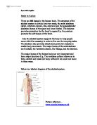
Parathyroid glands
Parathyroid glands are four small, oval-shaped glands that are embedded in the posterior surface of each lobe of the thyroid gland in the neck. The functions of the parathyroid glands are to secrete the hormone parathormone, (parathyrin PTH). The main function of PTH is to maintain and regulate the concentration of calcium in the blood. Precise calcium levels are important in the human body, since small deviations can cause muscle and nerve impairment. Parathyroid hormones stimulate the release of calcium by bones into the bloodstream, absorption of food by the intestines, and conservation of calcium by the kidneys. Calcitonin from the thyroid gland works in conjunction with PTH form the parathyroid glands to ensure essential activities such as blood clotting; nerve impulse transmission and muscle contraction can take place.
The Adrenal Glands
The two adrenal glands, which are also termed suprarenal glands, are small, triangular glands located on top of both kidneys. The glands are approximately four centimetres long and three centimetres thick and are situated on the upper pole of each kidney and enclosed in the renal fascia. An adrenal gland is made of two parts, the outer region is called the adrenal cortex and the inner region is called the adrenal medulla. The adrenal glands are stimulated by the hypothalamus releasing corticotrophin releasing hormones, and pituitary gland producing corticotrophin hormones. Both parts of the adrenal glands, the cortex and medulla, perform very separate functions.
The adrenal cortex produces three major groups of hormones from cholesterol, the glucocorticoids, mineralocorticoids and androgens, collectively known as the adrenocorticocoids. The main glucocorticoids include cortisol, (hydrocortisone), and corticosterone which are both secreted in response to stress and ACTH stimulus from the anterior pituitary gland. The effects of glucocorticoids on the body include the promotion of sodium and water reabsorption, regulation of carbohydrate metabolism, gluconeogenesis, and storage of glycogen. Cortisol, sometimes termed hydrocortisone, controls the body's use of fats, proteins, and carbohydrates. The other glucocorticoid, corticosterone, has the ability to suppress inflammatory reactions and also affects the immune system. In pharmacological quantities glucocoorticoids have an anti-inflammatory action, delay wound healing, suppress the response of tissues to injury and suppress the immune response. The main mineralocorticoid is Aldosterone. Aldosterone hormone inhibits the level of sodium and water excreted into the urine, ensures the correct amount of potassium is excreted, maintains blood volume, blood pressure and a general electrolyte balance in the body. When renal blood flow is reduced the kidney cells respond by releasing the enzyme renin, which in turn increases angiotensin production by the liver. Angiotensin causes the adrenal cortex to manufacture the hormone aldosterone that allows the kidney tubules to reabsorb sodium and water, which would increase overall blood volume and reduce renin production. This biological process is named the renin-angiotensin-aldosterone system and is one of many that helps maintain homeostasis. The last major group of adrenocorticocoids is the androgens or more commonly, sex hormones. Androgens produced by the adrenal cortex are believed to have little significance compared to those made by other glands. They are associated with deposition of protein in muscles and retention of nitrogen.
The adrenal medulla is composed from the same tissue as the sympathetic neurones and secretes hormones in response to sympathetic stimulation. This inner part of the adrenal gland is not essential to life, but greatly assists a person's ability to cope with physical and emotional stress. The two hormones secreted by the medulla are epinephrine, also called adrenaline, and norepinephrine a chemical also known as noradrenaline. Adrenaline is associated with the fight or flight response, and acts by priming the body for swift action. Adrenaline causes vasoconstriction in skin, vasodilatation of blood vessels concerned with the muscles, heart and brain, and causes pupils to dilate. Bronchioles in the lungs also dilate allowing an increase in oxygen consumption, metabolic rate increases and glycogenolysis is induced. Noradrenaline is a postganglionic chemical transmitter has little effect on smooth muscle and metabolic processes but has strong vasoconstrictive effects, thus increasing blood pressure.
Pancreatic islets
The pancreas is an elongated, tapered organ located across the back of the abdomen, behind the stomach. The right side of the organ, (the head), is the widest part of the organ and lies in the curve of the duodenum, the first division of the small intestine. The cells that form the islets are irregularly distributed through out the pancreas. There are three main types of cells that form the islets known as, alpha, beta and delta cells. Alpha cells secrete glucagon a substance that targets the hepatocytes to increase blood glucose levels and promote gluconeogenesis. The product of beta cells is insulin, a chemical that accentuates anabolic activities and regulates blood glucose levels. Insulin accelerates facilitated diffusion of glucose into cells, speeds up glycogenesis, speeds up lipogenesis, and increases protein synthesis and the uptake of amino acids. The hormone insulin also inhibits gylycogenolysis and gluconeogenesis. Increased blood glucose levels, amino acid levels and other pancreatic hormones such as gastrin, secretin and cholecytokinin stimulate secretion of insulin. Secretion is decreased by sympathetic stimulation, adrenaline, cortisol, and somatostatin, (GHRIH). The delta cells of the pancreatic islets produce GHRIH and several other hormones aimed at reducing the secretion of insulin and glucagon.
Pineal gland
The pineal gland or epiphysis cerebri, is a small, pea sized body that is reddish grey in colour. It can be located under the brain behind the third ventricle and is attached by a stalk to the upper portion of the thalamus. When the amount of light received through the retinas, (part of the eye), decreases the pineal body secretes more of a hormone named melatonin. Melatonin is a chemical messenger that is implicated in a wide range of human activities. It regulates circadian and dinurnal rhythms and effects the development of the sex organs before puberty and the menstrual cycle by igniting the secretions of gonadotrophins. Melatonin influences skin pigmentation, the immune system, calcium, sodium and potassium levels, and may defend against free radicals and help to slow down the aging process. The pineal gland hormone, melatonin, can prevent jet lag, is implicated in seasonal affective disorder, coordinates fertility, and allows for deep restful sleep patterns.
Thymus gland
The thymus gland is a primary lymphoid organ and lies in the upper part of the mediastinum behind the sternum and extends upwards into the root of the neck. It weighs about fifteen grams at birth and begins to grow until the individual reaches puberty when it begins to atrophy. Its maximum weight is around thirty-five grams and by the age of 40 it has returned to its weight at birth. The thymus consists of two lobes which are enclosed in fibrous capsules and that are composed of irregular branching frameworks of epithelial cells and lymphocytes. The thymus gland produces thymosin, a chemical that is required for the development of T- lymphocytes used in cell mediated immunity, (Tortora and Grabowski.2001). T- lymphocytes, a heterogeneous group of cells, are essential in protecting the body against invasions by foreign organisms and disease.
Gonads
A woman's two ovaries or reproductive glands are located on both sides of the uterus, below the opening of the fallopian tubes and lie in a shallow fossa on the lateral wall of the pelvis. The two ovaries are attached to the upper part of the uterus by the ovarian ligaments. The function of an ovary is to produce ova and secrete the hormones estrogen and progesterone, which affect many aspects of the female body including menstrual cycles and pregnancy. Oestrogen facilitates growth of the tissues of the sex organs and is responsible for the appearance of secondary female characteristics. Oestrogen also acts to strengthen bones and has a protective effect on the heart. Progesterone promotes the changes in the uterus that occur in preparation for the implantation of a fertilised ovum and prepares the mammary glands for milk production. Gonadotropic hormones produced by the pituitary gland control the levels of hormone secreted.
Testicles, (testes), are the male reproductive glands or gonads. Testes are egg shaped organs that are attached by spermatic cords and hang suspended in a pouch of skin named a scrotum outside the male body. The scrotum serves as a protective covering and serves to maintain the testicular temperature about 2 degrees below abdominal temperature. Two testes are found in the scrotum where they produce the male gametes, the spermatozoa, and the male hormone, testosterone. It is the anterior pituitary gland that releases LH, which stimulates the leydig cells of the testis to produce and secrete testosterone. Testosterone is responsible for the characteristics of the masculine body, including hair growth on the face and body and muscle development. Testosterone is essential for the production or sperm, spermatogenesis, and also acts to strengthen bones.
2.
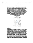
The digestive system is the collective name used to describe the alimentary canal, some accessory organs and a variety of digestive processes, which take place to prepare food eaten for absorption. The accessory digestive organs are the teeth, tongue, salivary glands, lingual glands, liver, gall bladder and pancreas. The process begins when food enters the digestive system through voluntary action via the mouth or oral cavity and the lips, (labia). The food is first processed by teeth, notably the anterior incisors that also work in partnership with the cheeks to prevent any falling out. Suitably sized portions are then retained in a closed mouth and chewed or masticated. This process involves teeth, the hard and soft palates, the movement of jaws, a tongue powered by intrinsic/extrinsic muscles to perform mechanical breakdown, plus some gland activity to initiate chemical breakdown. There are three pairs of salivary glands and some lingual glands, which open at various points into mouth, producing enzymes such as salivary amylase that initiates the chemical brake down of starch within a food. As a result of mastication the food is shaped against the hard palate to form a soft, flexible, easily swallowed mass named a bolus. In leaving the mouth the bolus of food must cross the respiratory tract, (trachea is anterior to oesophagus), by a complicated mechanism known as swallowing or deglutination, which empties the mouth and ensures that food does not enter the windpipe. Swallowing involves co-ordinated activity of tongue, soft palate pharynx and oesophagus. All of the processes up to now have been voluntary but become involuntary after reaching the pharynx. After travelling through the pharynx the bolus would arrive at the oesophagus or gullet. The oesophagus is a long muscular tube which terminates at the stomach and that is controlled at either end by sphincters. Food is assisted down the tube by mucus, gravity and the peristaltic movement. As the bolus continues through the lower oesophageal sphincter it enters the stomach, a J-shaped organ situated in the epigastric, umbilical, left hypochondriac region of the abdominal cavity, (Tortora and Grabowski.2001). The stomach produces gastric juices, which contain mucus for lubrication and protection, hydrochloric acid, gastrin, gastric lipase, intrinsic factor, and pepsin. Hydrochloric acid kills most bacteria and denatures proteins. Pepsin initiates the digestion of proteins and intrinsic factor aids the absorption of vitamin B12, which is essential for normal erythropoiesis. Gastric lipase enhances the digestion of triglycerides. When the bolus enters the stomach the muscular walls contort so that the food can churn and mix together with the gastric juices. The resulting substance is referred to as chyme, and is stored in the stomach for a short time until the creamy substance is voided via the pyloric sphincter to the duodenum by peristaltic movement. The First part of small intestine is the duodenum, a structure that is approximately twenty-five centimetres long and curves around head of pancreas. At its midpoint there is an opening, shared by the pancreatic duct and the common bile duct. Chemical degradation of small controlled amounts of food controlled by pyloric sphincter begins here, enzymes secreted by the pancreas and duodenum itself is aided by emulsifying bile. The pancreas delivers pancreatic juices containing pancreatic amylase, trypsin, chymotrypsin, elastase, carboxypeptidase, pancreatic lipase, and nucleases to the duodenum. The liver secretes bile salts and bicarbonate to neutralize the acidity of the chyme, absorb lipids and stimulate emulsification. The function of the gall bladder is to store mucin and concentrate bile for delivery to the first part of the small intestine. In the small intestine the chemical digestion of food is completed and most of the absorption of the nutrient materials takes place. The walls of the small intestine are covered with villi and microvilli, which serve to catch nutrients by providing an increased surface area and dispatch them into the rich venous and capillary drainage of the gut. The large intestine is the next part of the GI tract, its functions are to use bacteria such as escherichia, to brake down undigested carbohydrates, proteins, and amino acids, reabsorb water and then eliminate the drier residue as faeces. The large intestine has no villi, secretes its own mucus, is used for storage and can also synthesise vitamin B12 and vitamin K. Contractions of the intestinal wall create peristaltic motions which force the semi-solid waste contents into the sigmoid colon and rectum. The rectum terminates at the external anal sphincter which is under conscious control, (with exception of infants), through the pudendal nerve. The rectum wall contains stretch receptors, which initiate a defaecation reflex action that pushes the waste fectal material out of the body via the anus. The Anus has a voluntary and involuntary sphincter and the ability to distinguish whether its contents are in a gas or solid state. The waste material or faeces contain indigestible cellular plant and animal material, dead and live microbes, fatty acids, and mucosa.
3.
Minerals are inorganic elements that occur naturally in the Earths crust and that are essential for maintaining homeostasis within the human body.
a.
Potassium is the most commonly occurring intracellular cation and is involved in many chemical activities inside cells. It is important for muscle contraction, transmission of nerve impulses and maintenance of the electrolyte balance in the body. Although sodium and chloride are important, potassium is the most important dietary electrolyte. In addition to functioning as an electrolyte, potassium is essential for conversion of blood sugar into glycogen and the storage form of blood sugar in the muscles and liver. A potassium shortage results in lower levels of stored glycogen. Because exercising muscles use glycogen for energy, a potassium deficiency produces great fatigue and muscle weakness, the first signs of potassium deficiency. Potassium is important as it participates in the synthesis of protein from amino acids in the cell and is essential for normal growth, building muscle, heart function, muscle function, nerve function, and kidney and adrenal function. Deficiency of potassium is called hypokalemia, and symptoms include loss of appetite, muscle cramps, confusion and apathy. Hypokalemia is characterized by muscle weakness, slow reflexes, fatigue, mental confusion, irritability, weakness, heart disturbances, and problems in nerve conduction and muscle contraction. Other problems include dry skin or acne, insomnia, and loss of gastrointestinal tone. A sudden loss of potassium may lead to cardiac arrhythmias. Low potassium may impair glucose metabolism and lead to elevated blood sugar levels. In more severe potassium deficiency, there can be serious muscle weakness, bone fragility, central nervous system changes, decreased heart rate, and even death
b.
Calcium is associated with vitamin D, phosphorus and hardening of bones and teeth. It is essential for blood clotting, endocytosis, exocytosis, and normal nerve and muscle activity. The mineral is also important for chromosome movement, cellular motility, glycogen metabolism, and synthesis and release of neurotransmitters. Calcium supports normal blood cholesterol levels and overall heart health. A deficiency of calcium results in rickets in children and osteomalacia, both of which display a lack of bone mineralisation, (Tortora and Grabowski.2001). Calcium deficiency may also contribute to osteoporosis. A low level of calcium in the blood, (hypocalcaemia), makes the nervous system highly irritable and causes tetany, (spasms of the hands and feet), muscle cramps, abdominal cramps, and overly active reflexes.
c.
Iron is important for the formation of haemoglobin in red blood cells, it is also used in oxidation of carbohydrate. Iron is important in the transportation of oxygen from the lungs by way of the blood stream to the tissues. It is present in red blood cell protein, haemoglobin. A similar protein in muscle, myoglobin, also contains iron and stores oxygen for use during muscle contraction Deficiency of iron results in anaemia because iron is necessary to make haemoglobin, the key molecule in red blood cells responsible for the transport of oxygen. In iron deficiency anaemia, the red cells appear abnormal and are unusually small, (microcytic), and pale, (hypochromic). Deficiencies can cause Iron deficiency anaemia, heart palpitations, an impaired immune system, fatigue, hair loss, co-ordination problems, attention, learning and memory difficulties.
d.
Folic acid is essential for the production and maturation of erythrocytes. It is a vital component of enzyme systems synthesizing purines and pyrimidines built into DNA and RNA. A deficiency of folic acid would cause megaloblastic anaemia, abnormally large blood cells that would struggle to pass through capillaries. There is also a higher risk of neural tube defects, (NTDs), in babies born to folate-deficient mothers. The most common NTDs neural tube defects are spina bifida and anencephaly. Folic acid, also called folate or folacin, is one of the B vitamins, also known as B9. Your body needs it to produce red blood cells, as well as norepinephrine and seratonin two vital chemical components of the nervous system. Folic acid helps synthesize DNA and normalize brain function, and is a critical part of spinal fluid. Folic-acid deficiency most commonly causes folic-acid-deficiency anaemia. Symptoms include gastrointestinal problems, a red and sore tongue, cracks at the corners of the mouth, diarrhoea, and ulceration of the stomach and intestines. Other possible signs of folic acid deficiency include apathy, digestive disturbances, fatigue, greying hair, growth impairment, insomnia, laboured breathing, memory problems, paranoia, weakness, and birth defects in ones offspring.
4.
At birth the human skeleton is made up of 275 bones. As the body matures some of these bones, such as those in the wrists or ankles, fuse together leaving only 206 bones in the adult human body. Humans have an endoskeleton comprise of mostly hollow bones containing marrow cells inside. Ligaments connect bones to bones, and tendons connect bones to muscles. The Skeletal system includes bones, cartilage, ligaments, and tendons which all assist to determine the shape and symmetry of the body. There are six main functions of the skeletal system, support, protection, assistance in movement, mineral homeostasis, blood cell production and triglyceride storage.
There are two major systems of bones in the human body referred to as the axial skeleton and the appendicular skeleton. The axial skeleton is comprised of the 80 bones contained in the ribs, sternum, vertebral column, auditory ossicles, hyroid, and skull. The appendicular skeleton has 126 bones from the pectoral girdles, upper limbs, pelvic girdle, and lower limbs. Wolff's law suggests that the shape of a bone, to some extent, determines its main function and that conversely, the function may alter the shape, (Tortora and Grabowski.2001). Long bones such as the femur, tibia or fibula are slightly curved so that they may distribute and absorb stress evenly. They consist mostly of compact bone tissue in their diaphyses but also contain a large amount of spongy bone tissue in their epiphyses. Long bones are found in the lower extremities and their length and strength serve to support the weight of the body. Bones found in the upper extremities are termed short bones and are shorter and lighter. The function of short bone is to allow movement, provide elasticity, flexibility and to allow shock absorption. Examples of short bones include the carpal bones and the tarsal bones. Flat bones are generally thin and are composed of two nearly parallel plates of compact bone tissue. They provide attachment sites for muscles and give considerable protection to the vital organs. The skull, pelvis, scapula, ribs, and sternum are examples of flat bones. Twelve pairs of flat bones make up the rib basket, which serves to support the chest wall, and prevents it from collapsing, when the diaphragm contracts. Irregular bones have complex shapes and the amount of spongy or compact bone they are comprised from varies. The vertebrae are classified as irregular bones and their function is to provide protection for the spinal cord and central nervous system.
Apart from supplying the body with protection and support bones assist in movement. There are three main types of joints between two bones that allow different types of movement. A ball and socket joint consists of the ball like surface of one bone fitting into the cup like depression of another bone. Such joints are multiaxial because they permit movement around three axes plus all directions in-between. The coaxl joint, between the femur and pelvis, is a ball and socket and synovial joint. The cavity between the two bones contains the synovial membrane that produces synovial fluid, a liquid that acts as a lubricant. Both surfaces of each of the adjoining bones are also covered in soft, smooth cartilage, which acts as a shock absorber and allows movement. The tibiofemoral, or knee joint is an example of a hinge joint. Hinge joints are monoaxial and allow simple motion around a single axis. Pivot joining allows freedom of movement some where between ball and socket and hinge joining. An example of a pivot joint is the atlanto-axial joint, which allows an individual to turn their head from side to side, when wanting to produce the gesture 'no'. Other joints of the skull are extremely strong and mostly immovable. It is important to remember that a joint cannot function without a pair of opposing muscles to assist it. When one of the pair of muscles contracts, the other relaxes. The muscle performing the prime movement is the agonist, while the opposite acting muscle is the antagonist. An antagonist is never totally relaxed. Its function is to provide control and damping of movement by maintaining tone against the agonist, the whole process is termed eccentric movement.
The skeletal system also provides mineral homeostasis. Bone tissue stores several minerals, predominantly calcium and phosphorus, which contribute to the strength of bone, etc. Bone has the ability to release these minerals into the blood stream in order to maintain critical mineral balances. Bones also contain red bone marrow that is the production site for red and white blood cells, (hemopoiesis); both are vital for body function. As an individual matures some of the red bone marrow converts into yellow bone marrow. Yellow marrow consists primarily of adipocytes, and is used to store triglycerides.
Although human bones appear to be quite strong they can experience fractures and dislocations. A dislocation occurs when a bone is pushed out of its socket by a sudden unexpected pressure such as a fall. Dislocated bones often require a quick reverse pressure to realign them in the socket. Fractures are actual breaks in the bone and can be quite serious, taking anywhere from a month to six months to heal depending on the age and health of the individual and which bone is involved.
References
Mader. S, Sylvia. 2001. Biology. 7th edition. McGraw-Hill companies, Inc.
Wilson, Kathleen J.W., and Waugh, Anne. 1996. Ross and Wilson Anatomy and Physiology in Health and Illness. 8th edition. Churchill Livingstone.
Tortora, G.J., and Grabowski, S. 2001. Principles of anatomy and physiology. 9th edition. John Wiley & Sons, Inc.
Human and Social Biology
Assignment 2002
Eloise White