The Skeletal System
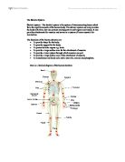
Skeletal system - The skeletal system is the system of interconnecting bones which form the rigid framework of the human body. The skeletal system not only provides the body with form, but also protects and supports its soft organs and tissues. It also provides attachments for muscles and serves as a system of levers essential for locomotion.
The functions of the human skeleton are:
* To provide shape for the body.
* To provide support for the body.
* To protect delicate organs e.g. brain.
* To provide a large surface area for the attachment of muscles.
* To provide a lever system through which muscles can pull.
* To provide a large surface area of the attachment of muscles.
* To manufacture red blood cells and to store fat, calcium and phosphate.
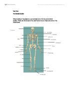
Here is a labelled diagram of the human skeleton:
The human skeleton is divided into appendicular skeleton and axial skeleton.
Axial Skeleton
Appendicula Skeleton
* Cranium
* Mandible
* Scapula
* Ribs
* Spinal column
* Pelvis
* Carpals
* Radius
* Ulna
* Humerus
* Pectoral girdle
* Femur
* Tibia
* Fibula
* Tarsal
Components of bones
Examples of each type of bone are shown below in diagram.
Bone is the hardest connective tissue in the body, mainly because it contains deposits of calcium phosphate and calcium carbonate. Bone acts as a store for calcium, and as a result of regular exercise more calcium is deposited, increasing bone density.
The bone matrix also contains collagen. Collagen gives the bone tissue a flexible strength, allowing it to cope with a certain amount of impact.
Hard, or compact, bone makes up the outer layer of all bones, giving them strength. Cancellous, or spongy, bone is typically found at the end of long bones. Cancellous bone is not as dense as hard bone because it contains cavities filled with bone marrow.
Bone marrow is soft tissue located in the cavities of the bones. The bone marrow is the source of all blood cells (Honeybourne et al. 2000)
Structure of typical bone
www.sirinet.net
Types of bones
There are different types of bones within the 206 bones which make up the skeletal system.
Long bones
These bones are cylindrical in shape and are found in the limbs of the body. These bones include:
* Femur
* Tibia
* Humerus
* Phalanges
The primary function of long bones is to act as levers, and they are therefore essential hen moving. Another vital function is the production of blood cells which occurs deep inside the bone.
Femur
Short bones
These bones are small and compact in nature, often equal in length and width. They are designed for strength and weight bearing, for example when performing a handstand, and include:
* The bones of the wrist (carpals)
* The ankle (tarsals) and calcaneum.
Tarsals
Flat bones
These bones offer protection to the internal organs of the body. Examples include:
* The sternum
* The bones of the cranium
* The bones of the pelvis
* Upon close inspection, it can be ...
This is a preview of the whole essay
Femur
Short bones
These bones are small and compact in nature, often equal in length and width. They are designed for strength and weight bearing, for example when performing a handstand, and include:
* The bones of the wrist (carpals)
* The ankle (tarsals) and calcaneum.
Tarsals
Flat bones
These bones offer protection to the internal organs of the body. Examples include:
* The sternum
* The bones of the cranium
* The bones of the pelvis
* Upon close inspection, it can be seen that the ribs are also flat.
They provide suitable sites for muscle attachment, with the origins of muscles often attaching to them. In this way the muscle contracting has a firm, immovable base against which to pull, and can therefore carry out its function effectively. The pelvis, sternum and cranium produce blood cells.
Scapula
Irregular bones
These bones are so named due to their complex, individual shapes and the difficulty in classifying them. They have a variety of functions which include protection. Examples include:
* The vertebrae (protect the spinal cord and help to absorb shock when running and jumping)
* The bones of the face
Vertebrae
Sesamoid bones
These bones have a specialised function: they ease joint movements and resist friction and compression. They are usually developed in tendons and are covered with a layer of articular cartilage as they exist where bones articulate.
Patella
Ligaments and Tendons
Structure
Function
Tendons
Round cord or band of connective tissue - non -elastic fibres
Joins muscle to bone - allows us to apply power and movement
Ligaments
White fibrous connective tissues - strong elastic fibres
Joins bone to bone - makes joint more stable
Achilles tendon Ligaments of the knee
www.footcaredirect.com www.steadman-hawkins.com
Structure of the vertebral column
The vertebrae column consists of 24 vertebrae and can be broken down into 5 different regions, each with its specific functions.
Cervical vertebrae (neck) - seven vertebrae make up this region. This area allows the head to move, such as 'nodding' and shaking, and also bending and twisting of the neck. This is where the muscles are attached.
Thoracic vertebrae (chest) - twelve vertebrae make up this area. These vertebrae are attached to the ribs and help to support the rib cage. There is some movement in this area allowing bending and turning of the trunk.
Lumbar vertebrae (lower back) - there are five vertebrae in this region and they are the largest of all vertebrae. This is where the back muscles are attached. This area allows the greatest amount of movement of bending forwards, backwards and side to side. This is also the most common area for back injuries due to the amount of movement and is an area which should be especially worked upon for flexibility exercises.
Sacral vertebrae - there are five vertebrae in this region which are fused together to become one. This is where the pelvis is joined to the spine and where the body mass is transmitted to the hips and legs.
Coccyx (tail) - there is four vertebrae fused together and is the base of the spine. It is all that remains of what was a tail before humans evolved, hence the name 'tail bone'.
The general functions of the spine are:
* To keep the body upright
* To help posture and movement
* To act as a shock absorber
* To protect the spinal column.
Joints
"The point of connection between two bones or elements of a skeleton (especially if the articulation allows motion)" www.cogsci.princeton.edu/cgi-bin/webwn
There are 3 main types of joints:
* Fibrous joints (or Sutured) - are very stable joints in which the bones are joined by very strong fibres allowing no observable movement e.g. the sutures in the skull (honeybourne et el. 2000)
www.science.ubc.ca
* Cartilaginous joints - are joined by a tough, fibrous cartilage which provides stability but also allowing a small degree of movement e.g. intervertebral disks
www.37c.com.cn/education
* Synovial joints - are the most common type of joint in the body, allow a wide range of movement in most cases although some (e.g. the sacro-lilac joint) are relatively immobile.
www.physicaltherapywebsites.com/library
There are 6 different types of Synovial joints; they can be un-axial (1 degree of movement), Bi-axial (2 degree of movement) or multi-axial (3 degree of movement):
. Hinge joint - is a uniaxial joint which only allows movement in one plane e.g. the knee joint only allows movement back and forth. Strong ligaments exist in order to prevent any sideways movement. www.marymount.k12.ny.us
2. Pivot joint - is also uniaxial, which allows rotation only e.g. the cervical vertebrae where the axis rotates on the atlas. www.marymount.k12.ny.us
3. Ball and socket - allows the widest range of movement and occurs where a rounded head of a bone fits into a cup-shaped cavity e.g. in the hip and shoulder. www.marymount.k12.ny.us
4. Gliding joint - is formed where flat surfaces glide past one another. Although mainly biaxial they may permit movement in all directions e.g. in the wrist, where the small carpal bones move against each other. www.marymount.k12.ny.us
5. Saddle joint - is biaxial and generally occurs where concave and convex surfaces meet e.g. the carpo-metacarpal joint of the thumb. www.sci.port.ac.uk/rad/ anatomy/05/006.htm
6. Ellipsoid joint - is biaxial, allowing movement in two planes e.g. the radio-carpal joint of the wrist allows back and forth as well as side to side movement. www.sci.port.ac.uk/rad/ anatomy/05/006.htm
Structure of a Synovial joint
All Synovial joints have several common features/characteristics. Here are these common features;
Cartilage - the bone ends forming the joints are covered in cartilage, which forms a smooth surface. This protects the bone tissue and reduces friction between bones, thus helping the smooth movement of the joint.
Joint capsule - this capsule is made of strong fibrous tissue. It surrounds the entire joint, adding extra stability, and encloses the joint completely, making it air tight.
Synovial membrane - This is a very thin membrane which lines the inside surface of the joint capsule. Its function is to produce Synovial fluid.
Synovial fluid - this is an oily liquid, yellowish in colour. It has several functions:
> It lubricates the cartilage surfaces by reducing friction between them. This permits even smoother movement.
> It forms a cushion of fluid between both bone surfaces, stopping the bone surfaces from grinding against each other. This reduces the wear and tear on the joint.
> It supplies nutrients for the cartilage
Ligaments - these are very strong bands of tough fibrous tissue which are fixed to the bones in a joint. They help to keep the joint in place, thus preventing dislocation.
Pads of fat - these can found in the gaps in and around the joint. They act as a shock absorber and cushion the joint.
Bursae - these are fluid-filled sacs. They prevent friction and wear and tear around the bone, ligaments and/or tendon, which can glide against one another.
<<<< SCAN PICTURE FROM PACK PICTURE >>>>>
Movement patterns of the body
To be able to name types of movement you must have a reference position of the body, this is called the anatomical position.
A person in the anatomical position is standing erect with the head, eyes and toes pointing forward, feet together with arms by the side. The palms of the hands are also point forward. (See the diagram on the below)
www.physio-net.com
When a person moves from the anatomical position: muscles, bones and joints work together to allow twisting, turning, bending, straightening and rotating. These movements are given special names;
Flexion -Bending the joint to make the angle between the two bones smaller. When you touch your right shoulder with your right hand, your elbow is in flexion (flexed).
Extension - Straightening a joint to make the angle larger. If you straighten your legs, the knees have undergone extension (extended).
Abduction - Moving away from the median plane. When you stand with your feet apart, your legs are in abduction (abducted).
Adduction - The opposite of abduction. If you sqeeze your knees together, you are adducting your legs.
Circumduction - A circular motion combining flexion, extension, abduction and adduction. Making circles in the air with your arms
Rotation - is the movement of bone around a central axis. For instance, the arm has both internal and external rotation.
Supination - A movement of the forearm in which the palm faces posteriorly. It is when your palms are faced up.
Pronation - A movement of the forearm in which the palm faces anteriorly. It is when your palms are faced down.
Eversion - is the movement of the sole of the foot outward at the ankle
Inversion - is the movement of the sole of the foot inward at the ankle.
Dorsiflexion - Movement of the ankle in the sagittal plane which decreases the angle between the foot and the lower leg when you point your foot towards your head.
Plantarflexion - Movement of the ankle in the sagittal plane which increases the angle between the foot and the lower leg when you stand on 'tip-toes' your ankles are in plantar flexion.