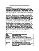
Mitosis can be simply described as having four stages-prophase, metaphase, anaphase and telophase. The stages follow one another without interruption. The entire mitotic process averages about sixty minutes, one hour, in duration, and the period between cell divisions, known as the interphase or interkinesis, can vary greatly but is considerably longer.
Interphase
In this stage the chromosomes are dispersed and appear as a network of filaments, long thin threads, called the chromatin. At some point prior to the prophase the chromosomes replicate themselves forming pairs of identical sister chromosomes, or chromatids; the deoxyribose nucleic acid, DNA, of the chromosomes is synthesized only during the interphase, not while mitosis is in process.
Prophase
During the prophase the two chromatids remain attached to one another at the region called the centromere, but each contracts into a compact tightly coiled body. The nucleolus and, in most cases, the nuclear envelope break down and disappear. During this time the spindle begins to form. In animal cells centrioles separate and move apart and radiating bundles of fibres, known as asters, appear around them. Some sets of fibre run from one centriole to the other; these are the spindle fibres. In pant cells the spindles form without centrioles.
Metaphase
Here the chromosomes congregate at a plane midway between the two ends to which the spindle tapers. This is known as the equatorial plane and marks the point where the whole cell will divide when nuclear division is completed. The ends of the spindle are the poles to which the chromatids will migrate. The chromatids are attached to the spindle fibres at the centromeres.
Anaphase
Here the spindle fibres join to the centromeres. Then the two chromatids of each chromosome separate and move to opposite poles, as if pulled along the spindle fibres by the centromeres. The separated chromatids are now chromosomes.
Telophase
In this stage new nuclear envelopes form around the two groups of daughter chromosomes, the new nucleoli begin to appear, and eventually, as the formation of the two daughter nuclei is completed, the spindle fibres disappear. The chromosomes uncoil to assume their dispersed distribution within the interfaced nucleus.
Cytokinesis
This process, which may begin before or after mitosis is completed, finally separates the two daughter nuclei into two new individual daughter cells.
A considerable variance in the degree and timing of these stages exists across species and cells can be classified by their mitotic characteristics. Despite the relative ease of observation of the physical stages of mitosis under a microscope, primarily because the chromosomes stain readily when in their coiled state, the exact chemical and kinetic nature of mitosis is not yet fully understood. For instance, the spindle has been determined to consist largely of thin, elongate tubules called microtubules, but their functions have yet to be understood.