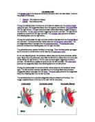
During the systole phase the right ventricle receives impulses from the and contracts. The atrioventricular valves close and the open. The de-oxygenated blood is pumped into the . The prevents the blood from flowing back into the right ventricle.
The pulmonary artery carries the blood to the lungs. There the blood picks up oxygen and is returned to the left atrium of the heart by the .
In the next diastole period, the semilunar valves close and the atrioventricular valves open. Blood from the pulmonary veins fills the left atrium. (Blood from the vena cava is also filling the right atrium.) The SA node contracts again triggering the atria to contract. The left atrium empties its contents into the left ventricle. The prevents the oxygenated blood from flowing back into the left atrium.
During the systole phase the atrioventricular valves close and the semilunar valves open. The left ventricle receives impulses from the Purkinje fibers and contracts. Oxygenated blood is pumped into the . The prevents the oxygenated blood from flowing back into the left ventricle.
The aorta branches out to provide oxygenated blood to all parts of the body. The oxygen depleted blood is returned to the heart via the vena cava.
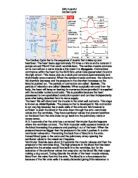
Atrial systole Ventricular systole Ventricular Diastole
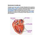
Cardiac conduction is the rate at which the heart conducts electrical impulses. Cardiac muscle cells contract spontaneously. These contractions are coordinated by the which is also referred to as the pacemaker of the heart. The SA node is composed of that has characteristics of both muscle and nervous tissue. The SA node is located in the upper wall of the right atrium. When the SA node contracts it generates nerve impulses that travel throughout the heart wall causing both atria to contract.
Another section of nodal tissue lies on the right side of the partition that divides the atria, near the bottom of the right atrium. It is called the . When the impulses reach the AV node they are delayed for about a tenth of a second. This delay allows the atria to contract and empty their contents first. The impulse then travels to the bottom of the ventricles along the AVN bumdle branches and to the Purkinje fibres causing the ventricles to contract from the bottom upwards forcing the blood out.