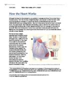
When the heart beats (systole) it pumps blood out of the heart, this contraction takes place in two stages. Firstly the left and right atria contract simultaneously causing blood to flow through the atrioventricular valves and into the left and right ventricles. Next the two ventricles contract, once again simultaneously, pumping blood out of the heart through the semilunar valves. The heart then relaxes (diastole) allowing it to once more fills up with blood. When you listen to the heart through a stethoscope you are likely to hear the heart, “lub-dub, lub-dub”. The first sound “lub” is not the contraction of the heart, as many believe it is, but purely vibrations caused by the closure of the atrioventricular valves (The tricuspid valve and the mitral valve). The “dub” sound is created on the same principle only that this time it is the pulmonary and aortic valve that close causing the sound vibrations. Naturally, the right side of the heart differs from the left side of the heart. The right side is responsible for receiving deoxygenated blood from the body and pumping it to the lungs where it is once more oxidized. The left side is then responsible for collecting newly oxygenated blood and pumping it out to the whole body, this is why the left side is much bigger and muscular, thus why most people assume that the heart is located left of the chest.
All blood enters the right atrium through two veins, the superior vena cava and the inferior vena cava. The superior vena cava collects the deoxidized blood from the upper body, and the inferior vena cava collects blood from the lower part of the body. Next the atria contract and the blood flows down through the tricuspid valve and into the right ventricle. Then the ventricles contract causing the deoxidized blood to travel through the pulmonary valve and into the pulmonary artery (the only artery in the body to carry deoxidized blood). The pulmonary artery eventually leads the blood to either of the lungs where it releases the non-useful and somewhat poisonous substance CO2 and receives fresh oxygen from the air outside of the body. Many people don’t know that the lungs primary job is not to respire but to control the pH level of the body through the process of breathing, by constantly releasing the potentially harmful gas (CO2).
The newly oxygenated blood re-enters the heart from via the left atrium through the pulmonary veins, and as odd as it may sound these are the only veins in the body to carry oxygenated blood. When the atria once again contract the blood is forced through mitral valve and into the very muscular left ventricle. Next the ventricles contract, pumping all of the oxygen rich blood out of the heart through the aortic valve and into the aorta, the largest artery in the body which eventually leads the blood to the cells of the body. The artery splits up and eventually becomes capillaries which will resemble a filter (permeable layer) only allowing the good and useful substances (food and oxygen) to be obtained by the cells. This process is called diffusion and the fluids that exit the bloodstream have no distinct colour, proving that the membrane only allows certains substances to pass and obviously haemoglobin isn’t one of them. Eventually the cells will send waste products such as CO2 into the bloodstream through veins which is how the blood becomes deoxygenated, any remaining fluid that isn’t obtained by the cells, ergo seeped all the way through tissue, is obtained by lymph’s and temporarily stored in a lymph node and eventually send back into the blood stream once again through veins. The blood travels up slowly and smoothly through the vein as there is very low pressure (some veins even have valves similar to the ones in our heart to prevent the blood from flowing back) and back to the heart through the superior and inferior vena cava. This process of blood flow is repeated countless times and does not stop till you die.
How does the heart actually beat automatically? The answer lies in a group of specialized cells found in the natural pacemaker of the heart, the sinoatrial node located near the right atrium. These cells have the ability to generate electrical activity automatically, separating charged particles and spontaneously send them into the left and right atria causing them to contract simultaneously. As mentioned the middle layer of the heart myocardium consists of cardiac muscle, a special kind of fibre not found anywhere else in the body with the ability to conduct electricity. These specialized fibres, present throughout the heart, will conduct the electrical impulse causing them to contract. From the atria the impulse travels to the atrioventricular node, between these two processes there is a natural pause allowing the ventricles to fill up with blood before moving on. From the atrioventricular node the impulse travels to the bundle of his, and eventually divides into the right and left bundle branches. The electrical impulse is spread quickly using purkinje fibres allowing the ventricles to contract at exactly the same time.
Although there is a specific pacemaker in the heart, technically any of the electrical tissues in the heart have the potential to function as one. However, the sinoatrial node generates an electrical impulse faster than any other electrical tissue in the heart. But should the pacemaker fail it will always have back-up, although the electrical impulse will generate at a slower rate. Although the pacemaker is in charge of creating an electrical impulse causing the heart to beat, specific nerves can manipulate the rate of which the cells generate electricity and the force of contractions (how powerful the heart contracts). These specific nerves are part of the autonomic nervous system, which consists of two sub-parts. The sympathetic nerves that is responsible for increasing the heart rate and increasing the force of contraction, and the parasympathetic nerves which do the opposite.
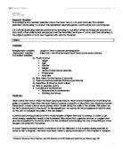
These specific processes all produce electrical waves we can measure, this is why it is called the bodies electrical system, or scientifically called cardioversion. The measurement is usually shown as a graph called an electrocardiogram (EKG). Here is an example of three heartbeats from an EKG.
http://health.howstuffworks.com/human-body/systems/circulatory/heart4.htm
Each part of the tracing has a lettered name:
- P wave – is the electrical activity that is passed through the atria and the beginning of its contraction.
- QRS complex – is spread of the electrical impulse through the ventricles causing them to contract.
- T wave – is the short natural pause that allow the ventricles to relax and the atria to fill up with blood.
The muscular wall of the heart, the myocardium, is constantly active (beats an average of 72 times a minute) and therefore needs a constant supply of oxygen and energy from blood. To provide this the heart has its own network of blood vessels known as the coronary arteries (the right and left), about four to five percent of the blood output of the heart goes to the coronary arteries. The two arteries branch from the main artery, the aorta, just after it leaves the heart, the left main coronary artery divides into the left anterior descending branch and the left circumflex arteries. Each artery supplies blood to different parts of the heart muscle and electrical system. The pattern of the coronary veins that collect waste products from the cells is very similar. Most of the blood collected in these specific veins is primarily collected by the coronary sinus, the large vein located at the back of the heart, which empties directly into the right atrium.
Heart disease or cardiopathy is the overall term for a variety of disease that affects the heart and its circulation. In 2007 it was the leading cause of death in the United States, England, Canada and Whales, accounting for whole 25.4% of the total deaths in America. Commonly, people suffer from high blood pressure. Some suffer heart disease as a result of diabetes. Others develop an arrhythmia, murmur, or irregular heartbeat. According to research women are becoming the greater half of the population to have heart disease, which might imply that they are more easily struck by it than men are. The large term heart disease can be split up into sub-groups, which include different types of diseases:
Coronary heart disease – The cardiac muscle, or myocardium, depends on a constant flow of blood, supplied by the coronary arteries. If this supply of blood becomes restricted in any way, the oxygen and nutrients (energy) will not be able to reach the heart muscle. A result of this could be a form of coronary heart disease (CHD).
-
Atherosclerosis – Atherosclerosis is caused by the narrowing as well as stiffening of the arteries due to fatty deposits known as atheroma accumulating in their walls.
- Angina – Chest pains that come with exertion and are relieved by rest, is a sign that the heart muscle isn’t getting an adequate supply of blood.
- Heart attack – A heart attack occurs when an area of cardiac muscle is not supplied with blood at all which means that it doesn’t receive any oxygen due to a blockage in an artery
Heart muscle disorders – The heart is mainly composed of cardiac muscle, or myocardium. Some heart disorders are caused by problems with this muscle or the sac-like membrane, the pericardium, which surrounds the heart. Severe heart muscle problems can eventually lead to heart failure, when the heart’s pumping power is reduced.
- Heart muscle disease – When the heart muscle becomes inflamed it is known as myocarditis, a variation is cardiomyopathy which is a non-inflammatory heart muscle disease.
- Pericarditis – This is when the pericardium becomes inflamed which is often a result of a viral infection or a heart attack.
- Heart failure – The hearts inability to effectively pump blood to the lungs and the body’s tissues.
Structural disorders – Structural heart disorders can affect people at any age. Congenital heart defects are present at birth, while valve disorders generally arise later in life. Structural disorders are usually with surgical techniques, for example diseased valves can be surgically widened or replaced.
- Congenital heart defects – Heart defects presents from birth, possibly due to a fault during early embryo development
- Valve disorders – There are several conditions that can affect the efficient functioning of any of the heart’s four valves
- Heart murmurs – Unusual heart sounds produced by turbulent blood flow which may be due to a heart valve defect
Circulatory and heart-rate disorders – A constant and adequate blood supply is essential for healthy tissues. If a blockage occurs in a blood vessel the tissues will be starved of oxygen causing the tissue damage and in severe cases lead to tissue death. The heart may also be affected if the electrical system is disturbed.
- Embolism – An embolus is a fragment of a material that breaks away from its original location which can cause partial or total blockage of a blood vessel
- Thrombosis – The partial or total blockage of an artery, vein or even the heart can occur whena blood clot (thrombus) forms due to a circulatory problem
- Aneurysm - Abnormal welling of a weakened arterial wall, which results in making the wall bulge out like a balloon
- Hypertension – Internal organs can become damaged if not treated due to a high blood pressure
- Arrhythmia – An abnormal heart rate of rhythm which is caused by a disturbance in the electrical system
So if you wish to keep your heart healthy for the rest of your life do following:
- Get plenty of exercise
- Follow a good diet
- Keep ypur heart clean and drug-free
“Be heart smart and have a healthy heart.”
Bibliography
Information (Internet Sources)
Information (Books, Text Books and School notes)
- The Human Body Book
- Concise Oxford English dictionary
- Biology Textbook
- School notes
Pictures/Diagrams/Graphs
“The average lifetime total heartbeats are based on an average of 72 beats per minute during an average lifespan of 75 years. 72 beats per minute x 60 = 4,320 beats per hour. 4,320 beats per hour x 24 = 103,680 beats per day. 103,680 beats per day x 365 = 37,843,200 beats per year. 37,843,200 beats per year x 75 = 2,838,240,000 beats in an average lifetime.”
http://health.howstuffworks.com/human-body/systems/circulatory/heart.htm
http://en.wikipedia.org/wiki/Heart
http://health.howstuffworks.com/human-body/systems/circulatory/heart5.htm
. National Center for Health Statistics.