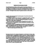
This is the linear sequence of amino acids:
Secondary structure is the ordered arrangement or conformation of amino acids in localized regions of a polypeptide or protein molecule. Hydrogen bonding plays an important role in stabilizing these folding patterns. The two main secondary structures are the alpha helix and the anti-parallel beta-pleated sheet. There are other periodic conformations, but the α-helix and β-pleated sheet are the most stable. A single polypeptide or protein may contain multiple secondary structures.
Both secondary structures give additional strength to proteins. The α-helix helps make though fibers like the protein in your nails, e.g. Keratin. The β-pleated sheet helps make the strength giving protein in silk, fibroin. Many proteins are made from both α-helix and β-pleated sheet.
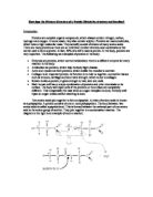
The polypeptides are held in position by hydrogen bonds. In both α-helices and β-pleated sheets the C=O of one amino acid to the H-N of an adjacent amino acid, like this, C=O---H-N.
Hydrogen Bonds
An α-helix is a tight, twisted strand; a β-pleated sheet is where a zigzag line of amino acids bonds with the next, and so on. This forms a sheet shape.
The protein shown, only achieves a secondary structure as the simple α-helix polypeptides do not undergo further folding. This is the structure of a fibrous protein. It is made of three α-helix polypeptides twisted together.
The tertiary structure of a polypeptide or protein is the three-dimensional arrangement of the atoms within a single polypeptide chain. For a polypeptide consisting of a single conformational folding pattern (e.g., an alpha helix only), the secondary and tertiary structure may be one and the same. Also, for a protein composed of a single polypeptide molecule, tertiary structure is the highest level of structure that is attained.
Tertiary structure is largely maintained by disulfide bonds. Disulfide bonds are formed between the side chains of by oxidation of two thiol groups (SH) to form a disulfide bond (S-S), also sometimes called a disulfide bridge.
This is due to the bending and twisting of the polypeptide helix into a compact structure.
Hydrogen bonds
Disulphide bonds maintain the tertiary structure
Ionic bonds
Quaternary structure is used to describe proteins composed of multiple subunits (multiple polypeptide molecules, each called a 'monomer'). Most proteins with a molecular weight greater than 50,000 consist of two or more non covalently-linked monomers. The arrangement of the monomers in the three-dimensional protein is the quaternary structure. The most common example used to illustrate quaternary structure is the hemoglobin protein. Hemoglobin's quaternary structure is the package of its monomeric subunits. Hemoglobin is composed of four monomers. There are two α--chains, each with 141 amino acids, and two β-chains, each with 146 amino acids. Because there are two different subunits, hemoglobin exhibits heteroquaternary structure. If all of the monomers in a protein are identical, there is homoquaternary structure.
Hydrophobic interaction is the main stabilizing force for subunits in quaternary structure. When a single monomer folds into a three-dimensional shape to expose its polar side chains to an aqueous environment and to shield its nonpolar side chains, there are still some hydrophobic sections on the exposed surface. Two or more monomers will assemble so that their exposed hydrophobic sections are in contact.
Some proteins do not have a quaternary structure. If they consist of just one folded polypeptide then they are classified as having tertiary structure. If they are simple fibers of α-helix or β-pleated sheets then they have only secondary structure.
This is a combination of a number of different polypeptide chains and associated non-protein groups to from large complex protein molecules.
Notes gathered from:
School notes,
Letts: Revise AS Biology and
Cambridge Advanced Sciences Biology 1