
Mitochondria and The Golgi Complex
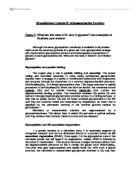
Work Book Section II a(i) Mitochondria a(ii) The structure and shape of the shape in the diagram suggested to me that it was a mitochondria cell. The structure of the all round shape and also the inner walls to the mitochondria cell. b(i) Golgi Complex b(ii) and 1c A membrane bound compartment in the interior of a cell. This compartment is involved in modifying, sorting and packaging lipid, carbohydrate and protein molecules for secretion or for delivery to other organelles. www.lsdn.com/glance_glossary.shtml One of the organelles that is in both the animal and the plant cells is the golgi apparatus. In this organelle, the endoplasmic reticulum (ER) sends vesicles(The function of the vesicles are to mainly transport proteins and other cellular material between cells and organelles) to the Golgi complex where they fuse with the cell membrane. Their membrane, which has now added to the membrane of the sacs of the golgi, empties it's contents of it into the golgi sac. Audesirk, Teresa; Audesirk, Gerald; "Fifth Edition Biology Life on Earth" Prentice-Hall; 1999 The Molecular Biology of The Cell. Second Edition. New York. Garland Publishing, Inc. 1989 d Single-membrane structure. The thickness of structure C shows to be only a single membrane cell. Also the structure when compared to other similar looking cells on the diagram, such as structure D, looks less rigid and

Skin Cancer
SKIN CANCER Skin cancer is the most common of all the types of cancers (Skin Cancer). It is a disease where malignant, or cancerous cells can be found in some layer of the skin. The epidermis, or the top layer of the kin, has three kinds of cells: basal cells, squamous cells, and the melanocytes (Skin Cancer). The melanocytes produce melanin, which is pigment that gives the skin its colour. Sometimes clusters of melanocytes can form growths called moles. These moles can sometimes become cancerous. Skin cancer tumours are formed when these cells excessively divide. They are known as malignant tumours. The tumours are not skin cancer when they are designated as benign. The cancer cells in the malignant tumours can break away and spread throughout the body. This can occur by way of the blood stream of lymphatic system (Melanoma). Skin cancer is categorized into three types: basal cell carcinoma, squamous cell carcinoma, and malignant melanoma. Basal cell carcinoma is, by far, the most common form of skin cancer (Skin Cancer). Luckily, it will never spread throughout the body. Basal cell skin cancer usually forms due to years and years of skin damage. Therefore, it is most often found on the sun-exposed areas of older adults. Such areas include the nose, face, back, and neck. Basal cell carcinoma is definitely curable with treatment. The second most common

WHAT IS LIFE?
WHAT IS LIFE? I hoped that the dictionary would offer some help in response to this cryptic question, but sadly it did not. The numerous dictionary definitions of life do very little to explain what life is and many people have varying opinions on what is and what is not alive. However I do I certainly agree with Antonio Lazcano who stated that "An all embracing, generally agreed upon definition of life has proven to be an elusive intellectual endeavour". So whilst it is unlikely that I will solve the question 'What is Life?' in this essay, I will attempt to explore some of the fundamental characteristics of life and the uncertain boundaries between what is alive and what is not. Many of us are taught that life is plainly defined by 'MRS GREN'; (Movement, Reproduction, Sensitivity, Growth, Respiration, Excretion and Nutrition); however this is a vast simplification of the topic. The answer to 'What is Life?' is unequivocally ambiguous which is highlighted by the distinct lack of scientific agreement. In addition, philosophers and theologians confuse the matter, contemplating over things such as robotic and computer life and the 'self-aware internet'. It seems that any attempts to define life are doomed to failure due to the simple fact that the transition from the complex organic molecules (which were existent between 1 and 2 billion years ago on earth) to primitive, living

Components of Biological Membranes.
Components of Biological Membranes Introduction. Biological membranes surround all living cells, and may also be found surrounding many of an eukaryotes organelles. The membrane is essential to the survival of a cell due to its diverse range of functions. There are general functions common to all membranes such as control of permeability, and then there are specialised functions that depend upon the cell type, such as conveyance of an action potential in neurones. However, despite the diversity of function, the structure of membranes is remarkably similar. All membranes are composed of lipid, protein and carbohydrate, but it is the ratio of these components that varies. For example the protein component may be as high as 80% in Erythrocytes, and as low as 18% in myelinated neurones. Alternately, the lipid component may be as high as 80% in myelinated neurones, and as low as 15% in skeletal muscle fibres. The initial model for membrane structure was proposed by Danielli and Davson in the late 1930s. They suggested that the plasma membrane consisted of a lipid bilayer coated on both sides by protein. In 1960, Michael Robertson proposed the Unit Membrane Hypothesis which suggests that all biological membranes -regardless of location- have a similar basic structure. This has been confirmed by research techniques. In the 1970s, Singer and Nicholson announced a modified version

Discuss how changes in control of the cell cycle contribute to cancer development Cancer is a multifarious disease, with a common feature that most tumours harbour one or more genetic mutations that allow them to advance outside their normal growth restr
Discuss how changes in control of the cell cycle contribute to cancer development Cancer is a multifarious disease, with a common feature that most tumours harbour one or more genetic mutations that allow them to advance outside their normal growth restraints. This proliferation is normally harnessed by the control of the cell division cycle, which in turn, is majorly regulated by the cyclin dependent kinases (Cdks) family of serine/threonine kinases and their regulatory partners, the cyclins (Errico, et al., 2009). In this essay, the roles of Cdks, cyclin complexes, regulatory proteins and other cell-cycle regulatory processes will be underlined, followed by an analysis of the genetic lesions in these regulators which may contribute to tumorigenesis. Fundamentally, cancer, or a neoplasm is a disease where cellular proliferation is no longer under normal growth control. The growth of this clone of cells exceeds, and is uncoordinated with that of normal surrounding tissues (NHS, 2009). Ultimately, this deregulation of growth and division of the cancer cells disrupts and interferes with the normal functioning of the body, either at its origin or through spreading to another location, eventually resulting in the potential death of the sufferer if left untreated. Other complex characteristics include the ability of the cancer cells to induce vascularisation of the tumour in
What are the roles of N- and O-glycans? Use examples to illustrate your answer.
Glycobiology tutorial II: oligosaccharide function Essay 2: What are the roles of N- and O-glycans? Use examples to illustrate your answer. Although the same glycosylation machinery is available to all proteins which enter the secretory pathway in a given cell, most glycoproteins emerge with characteristic glycosylation patterns and heterogeneous populations of glycans at each glycosylation site. What are the roles of these N- an O-linked glycans? Glycosylation and protein folding: The sugars play a role in protein folding and assembly. The proper folding and controlled assembly of many newly synthesized glycoproteins requires them to engage in a series of coordinated interactions with chaperones and enzymes through the attachment of a common oligosacchardide precursor, GlcNAc2Man9Glc3, to N-linked glycosylation sites. This sugar precursor is rapidly processed to GlcNAc2Man9Glc1 which can bind two lectins: the membrane bound calnexin (Clx) and its soluble homolog calreticulin (Clr). Lectins are oligosaccharide binding proteins. The interaction between Clx and/or Clr with nascent monoglycosylated glycoproteins provides access to a folding pathway. In their role as quality factors, Clx and Clr retain unfolded glycoproteins in the ER until they are correctly folded and assembled by chaperones, an event that is signalled by the permanent removal of the terminal glucose

Biological Membranes
Biological Membranes By Nishant Pradhan Ideas ) Biological membranes are a very complex part of a cell.- 2) Many variations of membranes exist even in a simple cell- 3) Give the main reason and importance of a cell membrane- 4) Different organelles, depending on their purpose have suitable membranes- 5) Give examples of different organelles having a specific type of membrane- 6) Explain the basic features of the plasma membrane- 7) Explain what pores do- 8) Explain why the membranes looks the way it does under the electron microscope- 9) Freeze fracture technique used to investigate inside the membrane- 0) Mitochondria having two separate membranes- 1) Temperature affects the membrane e.g. active transport may cease- 2) Plasma membrane in active transport- 3) Bounding cells together- 4) Chemical components of the membrane- 5) Gas exchange- 6) Cell membranes are made by the help of the golgi apparatus (explain)- 7) Chloroplast membranes- 8) Membrane around nucleus- 9) Cell wall shouldn't be confused with the cell membrane- 20) It is the cell membrane that is pulled during plasmolysis not the cell wall.- 21) A light microscope is not strong enough to clearly distinguish between the -features of a cell membrane, therefore an electron microscope is necessary PLAN ) Introduction - Cell membranes and the misconception. The complexity. 2) How the structure

The solubilisation and purification of an intrinsic membrane protein presents problems distinct from those encountered in purifying a conventional soluble protein. Discuss this statement.
The solubilisation and purification of an intrinsic membrane protein presents problems distinct from those encountered in purifying a conventional soluble protein. Discuss this statement. Word count: 1860 In order to answer the question, this essay will first describe how soluble proteins are purified. It will then describe the process of solubilising an integral membrane protein specifically, and demonstrate differences between the two processes. There are several methods for the purification of proteins in aqueous solution. Since these methods discriminate based on one characteristic that may be shared by several proteins, it is almost always necessary to use multiple methods to purify a protein from its cellular environment. First, the cell must be homogenised in order to make all the proteins within available. In theory, this presents a problem since proteins are mixed with proteases, and could be degraded. In practice vacuoles form spontaneously and quickly to mitigate this effect so it is not a problem that has to be contended with. After homogenisation, there are several chromatographic methods available to purify proteins completely. Size exclusion chromatography separates proteins based on molecular weight, as smaller proteins are retarded by the resin and so take longer to flow through. Ion exchange chromatography involves charged resin which binds charged amino
Macromolecular composition of a liver cell
Macromolecular Composition of the Liver cell Abstract A liver cell is to be homogenised and fractionated into a nuclei rich sediment and a nuclei free supernatant using centrifugation. After treatment with perchloric acid the samples are centrifuged producing supernatants containing glycogen, and these are decanted and stored. The sediments are washed, then treated with KOH and perchloric acid and centrifuged again. This supernatant contains ribonucleotides and it is also stored. The remaining precipitates are suspended in KOH and incubated to ensure it is fully dissolved. The addition of various reagents to each of the supernatants and suspended sediments will allow for an examination of the distribution of RNA, DNA, glycogen and protein, and for an explanation of why this is so. Introduction For supernatants to be produced for examination of this kind, the liver cells must be fractionated to allow specific organelles and molecules to be collected. This is done through homogenisation and differential centrifugation. During homogenisation citric acid is added and in put in a pre-cooled homogeniser; liver is easily broken up. It would be relatively much more difficult to homogenise a plant cell due to the presence of a cell wall, an outer layer that maintains cell shape and is made of cellulose, other polysaccharides and protein (Campbell and Reece, 2005). A centrifuge

Ribosomes; Structure and function.
Ribosomes; Structure and function 9 April 2003 Carly Brooks Ribosomes are cytoplasmic organelles discovered in both prokaryotic and eukaryotic organisms. Found in great abundance up to 10,000 in bacterial cells and many times more in eukaryotic cells, they comprise of proteins and rRNA molecules known as subunits, to form a large ribosomal complex. Both eukaryotic and prokaryotic ribosomes in association with transfer RNA (tRNA), act as a site for mRNA translation, assembling a specific sequence of amino acids into polypeptide chains, once the mRNA joins the two component subunits (large and small) of the ribosome. The tRNA is covalently bonded to an individual amino acid and has a complimentary nucleotide sequence, an anticodon, to each mRNA codon which form base pairs, adding specificity to the selection of the corresponding amino acids. The mRNA is linked by hydrogen bonds to the tRNA and is held in proximity to the amino acid so that a peptide bond is formed, this process occurs again and each amino acid is polymerized into a growing peptide chain. Ribosomes exist in two distinct forms; free and bound and may be positioned in several locations throughout the cell depending on cell function. Free ribosomes can occur individually, a monosome, or in clusters called polyribosomes or polysomes and are found in the