Influenza C
This is a common strain and causes mild illnesses in humans but not to the extent of causing an epidemic or pandemic.
Influenza B
Infectious only on humans and cause regional epidemics rather than pandemics
Often causes sporadic outbreaks of illness, especially in residential communities like nursing homes.
Influenza A
Responsible for regular outbreaks, including the one of 1918. Influenza A viruses also infect domestic animals pigs, seals, whales, horses, chickens, ducks) and some wild birds.
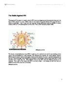
On the inner side of the influenza virion is an antigenic matrix protein lining. The influenza genome, which is organized into eight pieces of single-stranded RNA (A and B forms only; influenza C has 7 RNA segments). The RNA is packaged with nucleoprotein into a helical ribonucleoprotein form, with three polymerase peptides for each RNA segment.
Within the enveloped structure of influenza, there are also two varieties of spikes located. 80% of the spikes are identified as hemagglutinin (HA) and remaining 20% is known as neuraminidase (Na). These are formally known as glycoproteins.
There are currently 15 hemeagglutinin (Ha) and nine subtype that have been discovered within type A of influenza which are known as coded forms according to hemagglutinin and neuraminidase. H1N1, H1N2, H2N2 etc. The subtypes of influenza A virus are named according to their HA and NA surface proteins. For example, an H7N2 virus designates influenza A subtype that has an HA 7 protein and an NA 2 protein. Similarly an H5N1 virus has an HA 5 protein and an NA 1 protein.
Hemagglutinin is a primary protein responsible for binding to receptor sites on the cell membrane resulting in virion entrance within the cell It is so named because it causes agglutination which is a clumping of red blood cells. The red blood cell isn’t a primary host cell that the virus normally infects but has sialic acid on the membrane as the mucous membrane cells of the respiratory tract. This makes it a convenient cell type for assaying agglutination activity. Hemagglutinin is an integral membrane glycoprotein, shaped like a cylinder and is approximately 13.5 nm in length. It has three identical monomers that are constructed into a helix coil, consists of three heads (monomers) that contain the sialic acid binding sites. These monomers are later synthesized as precursors then broken down (glycosylated) into smaller polypeptides known as (HA1) and (HA 2) subunits. Each unit consists of a long helical chain anchored in the membrane by (HA2) and topped by a large (HA1) globule.
Hemagglutinin binds to the monosaccharide sialic acid which is present on the target cell surface. This binding induces cell membrane to engulf the virus and portion of the membrane to branch off forming a new membrane bounded compartment within the cell called an endosome. This endosome contains the engulfed virus. The cell then attempts to digest the contents of the endosome by the acidification of its interior transforming it into a lysosome. During endosome digestion, there is a decrease in pH to 6.0 therefore putting the Hemagglutinin molecule in an unstable form. This causes the molecule to unfold partially and release a peptide chain within the Hemagglutinin protein. This peptide chain is hydrophobic and acts like a fusion peptide by inserting itself into the endosomal membrane and locks on. The remains of the Hemagglutinin refolds into a new structure which is more stable at a low pH, the peptide is retracted and the endosomal membrane is drawn up to the virus membrane. The contents of the virus (RNA genome & proteins) are freed proceed into the cell cytoplasm and nucleus. Vital proteins set about to replicate the viral RNA strands by constructing a form of messenger RNA which is read out and translated into proteins by the cell protein making machinery.
The newly formed genes and proteins come together and bud as new viral particles coated with sialic acid which is unfortunate for the virus because this is the substance that binds the influenza virus to the cells for invasion. This results to unwanted particles such as antibodies binding to the sialic acid (like insects sticking to fly paper) therefore preventing further binding and infecting of cells. Thus antibody direction will neutralise the virus. This is the process by which influenza immunity is brought about during immunisation. However, the virus has an ace in the hole to countermeasure immunisation. This involves a catalytic reaction with an enzyme called neuraminidase.
This protein is a Glycoside hydrolase enzyme also known as a sialidase. There are four types of neuraminidase – Neu1 Neu2 Neu3 Neu4. Neuraminidase enzymes are found in a range of organisms. The most common neuraminidase is known as viral neuraminidase. Viral neuraminidase is used as an antigenic determinant found on the surface of the influenza virus. Its duty is to defend the virus by catalysing the hydrolysis of terminal sialic acid and residues from newly formed virions and from also host cell receptors that would otherwise block virus assembly process or virus particle incorporation. Neuraminidase also assists in the mobility of virus particles through the respiratory tract mucus bronchioles
It has been discovered that Neuraminidase is the enzyme that targets the sialic acid on the Hemagglutinin. Scientists are therefore trying to embrace the same methods through the production of drugs. However, the development of these new drugs become resistant to bacteria and viruses. The alteration of hemagglutinin is also under research in order to reduce the affinity or sialic acid thereby obviating the intervention of neuraminidase. The invention of these new mutants has shown very little to no signs of drug resistance. This results in the impaired binding between hemagglutinin and sialic acid reducing the infectivity of the strains by weakening their ability to dock with cells.
Bibiography:
Class Handout - W.Graeme Laver et al. Disarming Flu Viruses
BrockBiology of Microorganisms 12th ed pg- 563-564, 979