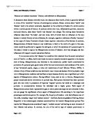
Figure 2: Depiction of flow cart elucidating the folding and misfolding of the protein (Adapted from David, 2003).
Most of the diseases are caused by tendency of aggregation unless the environment is tightly regulated. Random mutations act as another major cause for instability in the proteins causing interconversion between the two distinct forms of folded and misfolded structures. Alzheimer’s disease and neurological syndromes are some of the examples caused due to mutations leading to misfolding of the diseases (Dobson, 2004). Ageing also forms another devastating factor for protein aggregation and misfolding due to weakening of house keeping mechanisms leading to failure of protective schemes demanding more capacity of regulation which otherwise leads to many of the neurological and cardiac syndromes especially age related cardiac arrest and myopathies. The major factors contributing to this are mis regulation of chaperones and decline of degradatory mechanisms (Dobson, 2004). Many of the disorders like diabetes and other iatrogenic disorders like CJD (Prusiner, 1997) causing Amyloid deposition due to increase of serum levels decreasing the protective levels. Another factor contributing to misfolding of proteins is the lack of ability of the body to sustain the effect of heavy metals like mercury and cadmium on the human body causing mutations and deleterious effects on proteins (Dobson 2004) .
Table 1: Table depicting the proteins subjected to misfolding and diseases associated with them (Adapted from David, 2003).
Table 1: Some of the diseases caused due to protein misfolding termed as conformational diseases (Adapted from Dobson, 2001).
Evidences of relation between protein misfolding and disease:
1. Disease caused by neurodegenerative misfolding- Triosephosphate isomerase deficiency:
This is one of the neurodegenerative disorder mediated by the misfolding due to induced mutations. The result of the mutations causes various phenotypical exhibitions (Markus et al., 2006) Triosephosphate isomerase deficiency is one of the well known deficiency caused by glycolytic enzyme misfolding of proteins. As a result of this syndrome, haemolytic anaemia is caused in association with serious implication of another neurological disorder. Investigations revealed that the protein interconversion involves exchange of subunits between the oligomers which may cause decrease the normal biological functioning (Ferreira et al., 2001). In this neurodegenerative disorder, the mutation of these proteins caused by misfolded structures plays an important role. TPI, a glycolytic enzyme has a property of catalysing interconversion of D-glyceraldehyde 3- Phosphate to Dihydroxyacetone phosphate (DHAP). Deficiency of this enzyme causes haemolytic anaemia coupled with neurological dysfunction, causing death in children.
Low levels of TPI activity leads to metabolic blockage in glycolytic pathway which results in increased concentration of DHAP up to 50-60 fold. The blockage is increased in the presence of hetero-association of the mutant which causes reduction of catalytic activity by 10 fold. The pathway involves conversion of the metabolite to intermediates like Glu-6-PDH which is found in more levels when compared to the normal human beings which results in increased PPP in deficient cells. The pathology of these disorders is related to interconnection of both the pathways which result in reduction of TPI activity which is held responsible for normal ATP level regulation (Markus et al., 2006). TPI enzymopathy is one of the glycolytic enzyme deficiencies which are fixed along with neuro-degeneration which paves the pathway to many of serious neurodegenerative disorders like Alzheimer’s disease.
Figure 3: Depiction of cross talking of PPP with glycolysis (Adapted from Judit et al., 2002).
2. Protein misfolding in relation to cardiac diseases:
Investigations revealed that there are numerous degenerative disorders caused due to increase in misfolded pre amyloidogenic protein (POP). The research data reveals that PAO, pre Amyloid oligomers which is pre soluble is the major toxic entity responsible for pathology in Amyloid plagues and cardiac disorders. Many of the studies indicated the presence of PAO in many cardiomyocytes of samples in heart failure (Scott and Robbins, 2008). Experiments were conducted in transgenic mice to study the hypothesis of involvement of cardiomyocytes restriction enzyme in heart failure. The results of these experiments showed that polyglutamine residues of 83 (PQ83) or 19 (PQ19) were expressed by the mice confirming the accumulation of PAO in heart failure (Scott and Robbins, 2008). Further investigation suggests that long PQ lesser than 50 results in toxicity of neurological syndromes and shorter repeats of PQ tend to be benign. The expression of PQ83 results in accumulation of intracellular PAO which results in formation of aggregates and misfolded proteins causing death of cardiomyocytes leading to heart failure. Additional research proves that there is a presence of increased autophagy and necrosis associated with pathology of PQ83 cardiomyocytes. These investigations reveal the relationship between protein misfolding and cardiomyocytes death due to increase in intracellular concentration of PAO leading to heart failure (Scott and Robbins, 2008).
Many investigations in similar research lines suggest that autophagy as a critical pathway responsible for the deletion of aggregated and misfolded proteins and acts as a critical form for regulation of cell clearance. The deregulation and malfunctioning of autophagy is revealed as a most important contributing factor for pathogenesis of many cardiac and neurological disorders (Zhu et al., 2007). Autophagy is revealed as the most essential function in lysosomes and basal cardiomyocytes and up regulation and down regulation could lead to detrimental effects on heart (Zhu et al., 2007). Therefore all these studies reveal that there is a close relationship between homeostasis of heart and autophagy, misfolding of cardiomyocytes.
3. Protein misfolding in autoimmune disorders: Role of HLA-B27 in spondyloarthropathies
The investigation of misfolding caused in the antigen HLA-B27 in relation to spondyloarthropathies is yet to be published. The research work states that HLA-B27 (Human Leukocyte Antigen-B27) is major responsible factor causing spondyloarthropathies. The investigation focuses that the major reasons contributing to the pathology include presence of arthritogenic peptide, misfolding, presence of cell surface HLA-B27 dimers, indication of β2m (β2-microglobulin)deposition, indication of survival of microbes in the cells an β2m over expression (Chatzikyriakidou et al., 2011).
The hypothesis relating to protein misfolding suggests that the function of the antigen depends on the ability of its protein folding and simultaneous degradation of partially folded and misfolded proteins. Failure of degradation mechanisms or failure of protein folding ability along with gain of function processes like expansion of endoplasmic reticulum Amyloid fibrils, apoptosis, aggressive and inclusion bodies cause misfolding which result in progression of autoimmune disorders by immune related cells (Lopez, 2007). Misfolding of the processes is initiated by association of class I molecules in endoplasmic reticulum with β2-microglobulin (β2m) and antigenic peptides which are responsible expression of cell surface and presentation to the T-cells. HLA-B27 possesses an ability to form homodynes covalently linked by the cysteine-67 residues in α1 domain which causes excessive accumulation of HLA-B27 (Colbert et al., 2009). This excessive accumulation in the endoplasmic reticulum leads to activation of many nuclear factors like -κΒ (NF-κΒ) by unfolded response of proteins like UPR, resulting in release of pro-inflammatory cytokines like TNF-α, IL-1 and IL-6 causing inflammatory reactions (Chatzikyriakidou et al., 2011). Comparing to other HLA molecules, HLA-B27 tends to exhibit lesser folding rates which causes more stress in endoplasmic reticulum lading to exhibition of increased immune responses (Colbert et al., 2009).
4. Protein misfolding in Histone deacetylases (HDACs): Target of many neurodegenerative disorders
Histone deacetylases or lysine deacetylases (KDAC) play an important role as epigenetic regulators which act as catalysts in the removal of acetyl moieties from parts of lysine residues present in various proteins like histones. The role of HDAC’s is elucidated in various cellular functions like regulation of gene transcription and ability to regulate physiology of acetylated α-tubulin by initiating its capture (Li et al., 2011). Further, investigations also reveal that deacetylated α-tubulin plays an important role in response to misfolded and protein aggregation accumulation which serve as a major pathological syndrome in many neurodegenerative disorders like, Parkinson's, Huntington's and Alzheimer's diseases. The potential activities of α-tubulin deacetylases makes it as an important target for many diseases. HDAC6 plays an important role in degradation of protein complexes, formation of aggresomes and promotion of autophagy (Li et al., 2011).
HDAC 6 in aggresomes and autophagy: Research done on histone deacetylases reveal that HDAC6 initiates stress in the cellular environment which initiates the formation of protein aggregates from folded and misfolded proteins. Microtubule organizing unit then transports these aggregates through microtubule tracks which make use of dynein/dynactin motor complex. Later acetylation of α-tubulin takes place through HDAC6 which results in acetylation and association of dynein with α-tubulin making the transportation of protein aggregates easier by cystosol till aggresomes (depicted in figure 4) (Li et al., 2011) . Aggregated proteins are stored in the aggresomes where HDAC 6 initiates autophagic degradation. Later to the degradation, the fate of polyubiquitinated misfolded proteins is decided on the basis of balance exhibited between HDAC6 and the partner p97/valosin protein (VCP). HDAC6 additionally recruits proteins which ultimately lead to initiation of HSP90 dependent pathway which favors transcriptional process (Li et al., 2011).
HDAC6 in HSP chaperone system: HDAC6 also activates the shock response circuit against heat to elicit cellular responses, which leads to many cellular responses through chaperones. The mechanism of activity is attributed to the ability of chaperones to prevent formation of cellular aggregates by aiding refolding of the protein sequence or by catalyzing their delivery in degradation of ubiquitin-proteasome degradation system (Zhao, 2005). The active participation reduces the stress and safeguards the responses in stressed cells. HSP70, HSP90, HSP40 are several chaperones identified in this process which helps in regulation of various cellular responses (Li et al., 2011).
Figure 4 : Role of HDAC6 in aggresomes and autophagy (Figure adapted from Li et al., 2011).
5. Protein misfolding in NK-mediated cytotoxicity:
Investigations done in the recent past suggests that stress in the endoplasmic reticulum of thyroid cells through ER stressing agents, thapsigargin or tunicamycin initiate differentiation leading to the loss of phenotypic epithelial cells in experimental animals. The investigation was performed to examine the effect of stress induced in endoplasmic reticulum on the MHC class I to study the effect on function of natural killer cells in pathology of thyroid gland (Ulianich et al., 2011). Examinations in the laboratory performed on fetal thyroid cells revealed that thapsigargin and tunicamycin initiated the stress reaction on ER which subsequently reduced the cellular expression of MHC-1 plasma membrane, mainly through protein misfolding and unfolded protein response (UPR). The reduction of cellular response was accompanied along with reduction of natural killer cell activation and self tolerance which further increased the production and levels of INF-γ. The increased production of INF-γ caused increased activity of cytotoxicity against cells present in thyroid gland (Ulianich et al., 2011).
6. Effects of misfolding on glycation of albumin:
Glucose is a part of many vital body physiological functions and is necessary and vital nutrient responsible for cellular metabolism of oxygen. Protein modification and oxidative stress on the glucose leads to various disease states in the human body (Philippe and Emmanuel.,2011). One such damage noted is hyper glycemia and its associated damage caused due to increased glycation of albumin, which is circular protein present in the blood circulating system. Glycation of albumin is the main cause of impaired protein activity, protein unfolding and consecutive alteration in degradation mechanisms (Philippe and Emmanuel, 2011). All these activities lead to improper cellular functioning causing diabetic syndrome in the human body. Some other clinical implications caused due to the glycation of albumin by misfolding of the protein include neuropathy, nephropathy, retinopathy and coronary artery disease (Philippe and Emmanuel, 2011). Additional research is being undertaken of study the effect of glycation on diabetes and various other pathological progressions.
CONCLUSION:
Various investigations revealed that protein misfolding causes many diseases which are about 30 in number termed together as conformational diseases or misfolding diseases. The majority of conformational disorders are identified as a reason of ageing causing abnormal conditions of protein assembly and aggregation. Some of the various conformational disorders are caused due to deposition of various polypeptides like α synuclein, Amyloid β, tau and huntingten which leads to various neurodegenerative disorders of brain which are Parkinson’s disease and Alzheimer’s disorder. Gain of toxic function is another major disorder caused by protein deposits leading to diabetes type 2 through amylin and amyloidosis related to haemodialysis through β2 micro globulin. The research activities explain that the transmission of diseases is caused majorly due to the aggregation of proteins as in case of Creutzfeldt-Jacob disease (CJD), Bovine spongiform encephalopathy (BSE). There is also evidence that aggregates of Amyloid β, tau and Huntingten possess infectious properties. The review highlights various reasons for protein misfolding and the process of misfolding from normal protein folding mechanisms which is responsible for many of diseases affecting the human system like cardiac, pulmonary, neurodegenerative and systemic deficiencies. Many of the mechanisms of protein misfolding are under research on the basis of which therapeutic interventions could be framed to improve the health state of human beings. Recent evidences also show that the environmental and lifestyle modifications as a major cause of protein misfolding. These environmental factors could be controlled by planning appropriate lifestyle to reduce the deleterious effects of damage on protein functioning. Various ages related protein misfolding could be highly regulated by many preventive measures to improve protective mechanisms in the cellular metabolism (Markus et al., 2006).
The review emphasises the need of investigation in the reason for infection of the aggregated proteins and role of cellular factors like chaperone interactions in protein aggregation and clearance of aggregated proteins. There is a need of holistic approach in protein folding in order to understand the biophysical, biological and physiological mechanisms (Markus et al., 2006).
REFERENCES:
B. Hardesty and G. Kramer, Folding of a nascent peptide on the ribosome. Prog. Nucleic. Acid Res. Mol. Biol. 66 (2001), pp. 41–66.
B. Caughey and P.T. Lansbury, Jr., Protofibrils, pores, fibrils, and neurodegeneration: separating the responsible protein aggregates from the innocent bystanders. Annu. Rev. Neurosci. 26 (2003), pp. 267–298.
C.M. Dobson, The structural basis of protein folding and its links with human disease. Phil. Trans. R. Soc. Lond. B 356 (2001), pp. 133–145.
C.M. Dobson, Protein folding and disease: a view from the first Horizon symposium. Nature Rev. Drug Disc. 2 (2003), pp. 154–160.
Colbert RA, DeLay ML, Layh-Schmitt G, Sowders DP. HLA-B27 misfolding and
Spondyloarthropathies. Prion 2009; 3:15–26.
Chatzikyriakidou A, et al, What is the role of HLA-B27 in spondyloarthropathies? Autoimmun Rev (2011).
David R. Howlett (2003) Protein Misfolding in Disease: Cause or Response?Curr. Med. Chem. – Immun., Endoc. & Metab., Vol. 3, No. (4)377.
Dobson, C. (2004). Principles of protein folding, misfolding and aggregation. . 15(1). PP-3-16.
Ellis,R.J .protein folding:Importance of the Anfinsen cage. Current. Biol. 13 (2003), pp. R881–R883.
Ferreira, S. T. and De Felice, F. G. (2001). Theoretical model of prior propagation: A misfolded protein induces misfolding FEBS Lett. 498, pp. 129-134.( this reference is book, there is no title)
J. Scott Pattison and Jeffrey Robbins (2008) Protein Misfolding and Cardiac Disease:
Establishing Cause and Effect. Autophagy. 4(6): 821–823.
Judit Ola!h, Ferenc Orosz, Gyorgy M. Keseru, Zolta!n Kovari‹, Janos KovacsŒ, Susan Holla and Judit Ova!di (2002) Triosephosphate isomerase deficiency: a neurodegenerative misfolding disease. Biochemical Society Transactions,Volume 30, part 2
J.W.M. Höppener, M.G. Nieuwenhuis, T.M. Vroom, B. Ahrén and C.J.M. Lips, Role of Islet amyloid in Type 2 diabetes mellitus: consequence or cause?. Mol. Cell. Endocrin. 197 (2002), pp. 205–212.
Lopez de Castro JA. HLA-B27 and the pathogenesis of spondyloarthropathies.
Immunol Lett 2007;108:27–33.
Li G, et al, HDAC6 α-tubulin deacetylase: A potential therapeutic target in neurodegenerative diseases, J Neurol Sci (2011).
Markus.R, Heeren.G, Breitenbach.M, Lehrach.H, Krobitsch.S (2006) Triose Phosphate Isomerase Deficiency Is Caused by Altered Dimerization–Not Catalytic Inactivity–of the Mutant Enzymes. Available from www.plosone.org.
Muchowski PJ, Wacker JL. Modulation of neurodegeneration by molecular chaperones. Nat Rev Neurosci 2005;6:11–22.
NIA. (2010) Targeting Diseases Caused by Protein Misfolding or Misprocessing. Department of Health and Human Services( this is a pub med article, not a book).
Pepys MB. In: Weatherall DJ, Ledingham JG, Warrel DA, editors. The Oxford textbook of medicine, 3rd ed. Oxford: Oxford University Press; 1995. p. 1512–24.
Philippe Rondeau, Emmanuel Bourdon. (2011) The glycation of albumin: Structural and functional impacts. Biochimie (93) 645-658.
S. Prusiner, Prion diseases and the BSE crisis. Science 278 (1997), pp. 245–251.
L. Ulianich ,G. Terrazzano, M. Annunziatella,, G. Ruggiero, F. Beguinot, B. Di Jeso. (2011) ER stress impairs MHC Class I surface expression and increases susceptibility ofthyroid cells to NK-mediated cytotoxicity. Biochimica et Biophysica Acta 1812.431–438
Vendruscolo, M., Zurdo, J., MacPhee, C. E. & Dobson, C. M. Protein folding and misfolding: a paradigm of self-assembly and regulation in complex biological systems. Phil. Trans. R. Soc. Lond. 361, 1205–1222 (2003).
Zhao R, Houry WA. Hsp90: a chaperone for protein folding and gene regulation.
Biochem Cell Biol 2005;83(6):703–10.
Zhu H, Tannous P, Johnstone JL, Kong Y, Shelton JM, Richardson JA, Le V, Levine B, Rothermel BA, Hill JA. Cardiac autophagy is a maladaptive response to hemodynamic stress. J Clin Invest 2007;117:1782–1793.