Filtration: filtering the blood of its small molecules and ions.
Reabsorption: reclaiming the needed amounts of useful materials.
Secretion: surplus or waste molecules and ions are left to flow out as urine (5).
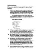
Blood enters the kidney through the renal artery. The renal artery then branches into interlobar arteries. These further branch into interlobular arteries. After the blood has entered the glomerulus through the afferent arteriole it leaves by the efferent arteriole to circulate the network of capillaries
that surround the nephron. The glomerulus is composed of three layers. The endothelium contains tiny pores or fenestrae that allows all solutes in blood plasma to leave the capillaries and prevents blood cells and platelets passing through. The basal lamina is composed of collagen fibres and proteoglycan, laminin, and other glycoproteins. The collagen is produced by the epithelial cells and forms a lattice like structure. It acts as a selective barrier by preventing the passage of larger molecules. (Tortora). Water, small molecules (glucose, amino acids, urea), electrolytes (sodium, chloride, potassium) some small proteins pass through the glomerulus while blood cells and large plasma proteins are retained in the blood stream. This procedure is called filtration. The filtered liquid, glommerular filtrate collects in the Bowman’s capsule and flows into the proximal convoluted tubule, PCT. The filtrate enters the tubule at 105-125ml/min. In the PCT 100% of glucose and amino acids are recovered by active transport; 80-90% of bicarbonate ions (HCO3-) are also reabsorbed along with 65% of water, sodium (Na+) and potassium (K+); 50% of chloride (CL-) and some calcium (Ca2+), magnesium (Mg2) and phosphate (HPO42) reabsorbed. These solutes are very valuable to the body, therefore are saved and reabsorbed into the blood supply. There are two types of transporters in this area. One reabsorbs Na+ ions while the secondary active transport process; Na+/ H+ antiporters reabsorb Na+ while secreting hydrogen (H+) ions at the same time. By the time the filtrate has travelled through the PCT, all the materials that need to be reabsorbed, have been reabsorbed. The filtrate that enters the descending limb of the loop of Henle, (LOH) enters at 40-45 ml/min. This is much less than when it entered the PCT. Here, the mixture is very different because there is no longer any glucose, amino acids or nutrients. The osmolarity of the tubular fluid is similar to that of blood.
Reabsorption is still taking place in the LOH. 15% of the water is reabsorbed and 20-30% of Na+, K+ and Ca2+; 10-20% of HCO3- and 35% CL-. All the water absorbed in the LOH does so in the descending limb, no water is absorbed in the ascending limb as it differs in permeability. The descending limb is permeable to water and the ascending limb is permeable to sodium ions and chloride ions. The differences enable more tubular reabsorption to occur. The loop is mostly made of simple squamous epithelium, which are slightly fatter than usual but thinner than regular cuboidal epithelium. The epithelium of the descending limb does not allow solutes to pass through because the cells don’t have the protein channels here. In the ascending limb, sodium is actively secreted out into the medullary interstitial fluid.
The fluid enters the distal convoluted tubule (DCT) at the rate of 25ml per minute. The amount of fluid is considerably lower here than in the PCT and the LOH because about 80% of the filtered water has already been reabsorbed back into the blood supply. Na+ and Cl- are still being reabsorbed by symporters under the control of aldosterone. Aldosterone stimulates sodium reabsorption in the
DCT and collecting ducts but is more important for controlling blood potassium levels. If too much potassium is excreted, hypokalemia, manifested by muscle weakness and possibly heart fibrillation could occur. The ions themselves regulate aldosterone secretion; increased blood potassium or decreased blood sodium levels stimulate the secretion of aldosterone from the adrenal cortex. As the filtrate leaves the DCT, the volume will be reduced but it will be isotonic rather than hypotonic.
After going through the distal tubule, the urine passes into the collecting ducts. When the filtrate has reached the collecting duct, 90-95% of the filtered solutes and water have been reabsorbed back into the blood system. The absorbed water is returned to the circulatory system via small capillaries called vasa recta. These are arranged in a tangle around the LOH, PCT and the DCT within the medulla. The collecting ducts are water permeable therefore the huge concentration gradient between the medulla and the collecting ducts remove water out of the urine into the medulla. This has produced concentrated urine. The extent of how concentrate the urine is depends on how water permeable the collecting ducts are. The water permeability of the collecting ducts is controlled by anti-diuretic hormone (ADH). In the absence of ADH, the collecting ducts are water impermeable, meaning no water is reabsorbed and a large volume of dilute urine is produced. In the presence of a high concentration of ADH the collecting ducts are highly water permeable, a lot of water is reabsorbed and a small volume of very concentrated urine is produced. Urine collects in the renal pelvis and then flows to the bladder. It does this 24 hours per day; fortunately the urine can be stored in the bladder until the time comes.
Renal failure of the kidneys occurs for various reasons. Causes include surgery or trauma, pregnancy, various medical conditions, nephrotoxins, or irreversible conditions that diminish nephron function. If the kidneys are damaged to such an extent that it can’t function correctly, the blood has to be filtered and cleaned artificially. This process is called Dialysis. Without dialysis, toxic wastes build up in the blood and tissues, and cannot be filtered out by the kidneys. This condition is known as uremia, which means "urine in the blood." The build up of waste may eventually lead to death. There are two types of Dialysis, Hemodialysis uses a dialyzer, or special filter, to clean your blood. The dialyzer connects to a machine where blood travels through tubes into the dialyzer. The dialyzer filters out wastes and extra fluids. Then the cleansed blood flows through another set of tubes and back into your body. Peritoneal works similar to hemodialysis but the blood is cleaned inside the body rather than through a machine. The abdomen has a peritoneal cavity which is lined by a membrane, called the peritoneum. The peritoneal cavity is filled with dialysis fluid that enters the body through a permanently implanted catheter. Excess water and wastes pass though the peritoneum into the dialysis fluid. This fluid is then drained from the body and disposed of.
Appendix 1.
Functions of the kidney
References
http://users.erols.com/jkimball.ma.ultranet/BiologyPages/K/Kidney.html#The_Kidney_and_Homeostasis
http://www.e-kidneys.net/
http://www.health.herts.ac.uk/depts/postreg/paulineF/renalPhysiologyRevision.html
http://science.howstuffworks.com/kidney.htm
http://dtc.pima.edu/biology/202/les10/lesson10b.pdf
http://courses.smsu.edu/emh420f/unit_5/Unit%205F/Unit%205F-1/Unit%205F-1a/Unit%205F-1a%20Internal%20Structure.htm
Tortora and Grabowski, 2003, Principles of anatomy and physiology, Wiley and sons Inc.
John Clancy and Andrew J Mcvicar,1995, Physiology and Anatomy a homeostatic approach