Urine passes out into the pelvis before it passes down the ureter.
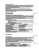
The nephron is the functional unit of the kidney. At one end of the nephron is the cup-shaped Bowman’s capsule. Immediately below the capsule is a twisted region called the proximal convoluted tubule. This leads into the long, hairpin-like loop of Henle, which runs deep into the medulla and then back out of the cortex, where it forms another twisted region called the distal convoluted tubule. This is joined to a collecting duct, which carries urine through the medulla to the pelvis of the kidney.
Glomerular ultrafiltration:
Each Bowman’s capsule is supplied with blood by an afferent arteriole. It branches inside the Bowman’s capsule to form a knot of capillaries, the glomerulus. These join up again to form the efferent arteriole, which take blood away from the Bowman’s capsule. The afferent arteriole is much wider than the efferent arteriole; this means that pressure is built up.
Ultrafiltration involves the filtering (under pressure) of small molecules out of the blood and into the Bowman’s capsule. The blood entering the glomerulus is separated from the space inside the Bowman’s capsule, by two cell layers and a basement membrane.
- The first layer is the endothelium of the capillary. In the glomerulus, this single layer of cells has thousands of gaps.
- The basement membrane between the two cell layers is composed of glycoprotein and collagen fibres. Its mesh-like structure acts as the filter during ultrafiltration.
- The second layer makes up the wall of the Bowman’s capsule. These cells have many tiny finger-like projections, with gaps in between them and are called podocytes.
The gaps in the capillary endothelium and the Bowman’s capsule wall allow most molecules through. But the basement membrane prevents large molecules (proteins and blood cells) from passing through and so acts as the filter. Only small, soluble molecules can pass through the basement membrane. The rate at which fluid seeps from the blood in the glomerular capillaries into the Bowman’s capsule depends on the difference in water potential between the contents of the two areas. The water potential of the blood plasma in the glomerulus is higher than that of the liquid in the Bowman’s capsule. Overall the effect of differences in pressure outweighs the effect of differences in solute concentration.
This filtration is a passive process, because molecules are not actively transported across the membranes, they diffuse down a concentration gradient as well as being pushed through due to the pressure build up in the glomerulus. The membranes are non selective.
Reabsorption in the proximal convoluted tubule and secretion into the distal tubule:
Many of the substances in the filtrate need to be kept by the body, so they are reabsorbed into the blood as the fluid passes along the nephron. This is called selective reabsorption since only certain molecules are reabsorbed. The structure of the cells making up the wall of the proximal convoluted tubule has many of the adaptations associated with active transport:
- Microvilli provide a large surface area for absorption.
- Numerous mitochondria provide ATP for active transport.
Substances do not just diffuse freely through the membrane, they can only enter through carrier proteins in the membrane. This is facilitated diffusion. All this results in the surrounding blood having a relatively high solute concentration. So a large amount of water passes out of the filtrate in the proximal convoluted tubule, back into the blood by osmosis.
Reabsorption of water in the loop of Henle and collecting duct:
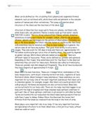
The loop of Henle is a hairpin loop that runs deep into the medulla and then turns and goes back to the cortex again. The function of the loop of Henle is to create an area of high solute concentration deep in the medulla. The collecting duct of each nephron pass through this area and so a lot of water can be reabsorbed from the collecting ducts by osmosis. Concentrated urine can be produced as a result. The ascending limb is more permeable to salts and less permeable to water. As the filtrate moves up, sodium and chloride ions move out passively at first and are actively pumped out of into the surrounding tissue. This causes water to pass out of the descending limb by osmosis. As a result the filtrate becomes more concentrated as it passes down the descending limb of the loop. The net result is that the solution concentration at any part of the loop is lower in the ascending limb than it is in the descending limb. As the collecting ducts pass through the medulla to the pelvis, they pass through this region of high solute concentration. So water is drawn out of the collecting ducts by osmosis into surrounding capillaries’ blood, resulting in a far more concentrated urine.
Control of water reabsorption:
Osmoregulation is the homeostatic control of body water, which is operated on a principle of negative feedback. The receptors responsible for detecting changes are located in the hypothalamus of the brain. These osmoreceptors react to change in the solute concentration of the blood as it flows through the hypothalamus. It detects if blood has a low water potential (more concentrated) and stimulates the pituitary to release antidiuretic hormone (ADH). The release of ADH into the bloodstream brings about the following:
- ADH make the distil convoluted tubule and the collecting duct more permeable to water.
- This allows more water to be reabsorbed from the distal convoluted tubule and the collecting duct into the region of high solute concentration in the medulla.
- This produces a smaller volume of more concentrated urine.
If the blood has a high water potential (less concentrated), it is detected and less ADH is secreted by the pituitary. This decrease in the amount of ADH in the bloodstream result in the following:
- The distil convoluted tubule and the collecting duct becomes less permeable to water.
- Less water is reabsorbed into the medulla.
- Larger quantities of dilute urine are produced.