Secondary structures of proteins
Alpha helix Beta pleated sheet
©Brannon Carter and Robin L. Carter
The three-dimensional shape of a protein is known as a tertiary structure. This is a specific shape which is held together by weak bonds between the R groups of the amino acids in the chain. The specific shape needed by a protein in order to carry out it’s particular function is created by the tertiary structure. It is essential that enzymes have a specific shape to enable them to catalyse particular reactions.
The tertiary structure of a protein.
©Brannon Carter and Robin L. Carter
Globular and fibrous are the two major types of protein. Fibrous proteins are fibers or sheets constructed by the joining of polypeptides. These are insoluble in water and strong, which makes them suitable structural proteins. An example of a fibrous protein is Keratin, which can be found in hair and fingernails. Skin, bone, and blood vessels also contain the fibrous protein collagen.
Globular proteins tend to have a biochemical function, rather than a structural function, as they are soluble in water, and have an approximately spherical shape. An example of a globular proteins is an enzyme.
The lock and key hypothesis
Enzymes are constantly moving in random motion. When the enzyme and a substrate molecule collide, the substrate binds to the enzyme’s active site, creating an enzyme-substrate complex. A product is formed with the reaction of the substrate inside the enzyme-substrate complex, and then it leaves the active site. The enzyme is capable of reacting again with other substrate molecules, as it is unchanged in the reaction. The lock and key mechanism describes how the enzyme catalyses a reaction. The enzyme is the “lock”, and the “key” is the substrate. The diagram shows how the enzyme is joined by the substrate.
Enzyme Substrate
©James K. Hardy and the University of Akron.
Induced fit hypothesis
The only way in which the enzyme-substrate complex can be formed, is if the substrate and the enzyme’s active site have complementary. It has been discovered that the substrate and enzyme are only fully complementary when the substrate is “bound” to the active site.
The diagram shows that the shape of the substrate is similar to that of the active site, but they are not fully complementary. The enzyme will change shape when the substrate binds to the active site, to make the enzyme and substrate fully complementary. The substrate is now able to join the active site and react to produce the product.
©James K. Hardy and the University of Akron.
Enzyme Substrate
Activation energy
Activation energy is the minimum amount of energy required for a reaction to take place.
Not all collisions will result in a reaction. The only way in which a reaction can occur, is when a collision takes place with sufficient energy between the reacting substances to overcome the energy barrier. Sufficient energy is created if the substances are traveling fast enough, and in the correct direction at a collision. An increase in the number of collisions with enough energy results in an increase in the speed of the reaction.
The energy barrier can be overcome by heating the reacting mixture, as the molecules will move faster when more heat is added. The name of the movement energy of the molecules is the kinetic energy. The likelihood of a collision taking place is increased when the amount of kinetic energy is increased. The human body has a temperature of 37 0C, and most of the substrate molecules do not have sufficient energy to overcome the energy barrier when a collision takes place, but increasing the temperature would lead to the chemicals, such as proteins, becoming damaged.
The enzymes inside the body allow reactions to take place at 37 0C. Activation energy is defined as the minimum amount of energy required for a reaction to occur. The enzymes lower this activation, making it easier for a reaction to take place. The activation energy is also lowered by the formation of the enzyme-substrate complex.
A graph showing the effect of enzymes on reaction rate.
©James K. Hardy and the University of Akron.
Co factors
A number of enzymes will only carryout a reaction in the presence of a non-protein substance known as a co-factor, which binds to the active site, and can aid a chemical reaction taking place by acting as an energy source.
There are three known types of co-factor:
- Inorganic ions, such as metal ions, are capable of acting as co-factors. These reduce the activation energy by joining to the enzyme or substrate, and are known as activators, which are believed to create the enzyme-substrate complex more easily. An example is the action of amylase on starch, which occurs more quickly in the presence of chloride ions.
- A coenzyme is also a co-factor, which will momentarily join to the active site of the enzyme and take part in the reaction. A link between two different reactions can be made with a coenzyme.
- A coenzyme that is permanently joined to the enzyme by a covalent bond is called a prosthetic group. This assists the enzyme to act as a catalyst.
Enzyme
Cofactor
Substrate
©James K. Hardy and the University of Akron.
The effect of temperature on the rate of reaction
The graph indicates the effect that temperature has on reaction rate. Due to the low temperature, and therefore kinetic energy, then enzyme and substrate at A travel slowly. This results in a slow rate of reaction as there are very few successful collisions between the molecules.
An increase in temperature will provide the enzyme and substrate molecules with more kinetic energy, which will result in an increased number of successful collisions between the molecules. The number of enzyme-substrate complexes will also increase with the reaction rate. It is these factors that will determine the rate of reaction. For these reasons, the rate of reaction will increase with the temperature, as shown by the graph.
The substrate and enzyme molecules will have a large amount of kinetic energy at a high temperature. Molecules vibrate as a result of kinetic energy. Therefore, an increase in temperature will cause the molecules to vibrate more. The shape of the enzyme will change if the hydrogen bonds are broken, i.e. the enzyme will denature. This will happen if the molecules have a large amount vibrational energy. By observing the graph, denaturing of the enzyme is taking place between points B and C. The rate of reaction will decrease beyond point B if there is an increase in temperature as more of the enzyme is denatured, which means that enzyme-substrate complexes are unable to be created. The consequence of this is that no more product can be formed.
The graph indicates that the maximum rate of reaction is at point B. The maximum rate at which the product is being formed is at this temperature. This temperature is known as the optimum temperature. Enzymes have an optimum temperature between 40 0C and 50 0C.
© Lea, Lowrie, McGuigan
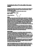
The effect of PH on reaction rate
A large number of cells have a PH of 7, which is also the optimum PH of almost all enzymes. The PH of extracellular (outside of the cell) enzymes is different to that of enzymes inside cells.
PH is a measure of the concentration of hydrogen ions (H+), or the number of hydrogen ions in a certain volume. Hydrogen bonds and ionic bonds are affected by the concentration of H+ ions. The enzyme’s specific three-dimensional shape can be altered by a change in hydrogen bonds. The enzyme will denature when it’s shape changes.
The graph displays the effect on an enzyme-catalysed reaction. The Ph at point A is low, therefore it is a strong acid, which affects the number of hydrogen and ionic bonds in the enzyme. The shape of the enzyme will be altered, causing it to denature.
There is a high PH at point C, therefore it is a strong alkaline. The hydrogen and ionic bonds are again affected by the high PH, therefore the enzyme will also denature.
© Lea, Lowrie, McGuigan
The graph indicates that an increase in PH between points A and B causes the rate of reaction increase. The PH level between points B and C also increases, but the rate of reaction decreases. Therefore, the maximum rate of reaction is at point B, indicating that the enzyme has an optimum PH of seven.
The effect of substrate on the reaction rate
The graph displays the effect that the amount of substrate has on reaction rate in an enzyme controlled reaction. Point A shows a low amount of substrate, which means that a small number of enzyme-substrate complexes will be produced, causing a low amount of product to be created, indicating that the rate of reaction is low. The graph shows that the reaction rate increases between points B and C as the number of collisions between the molecules of enzyme and substrate is increased.
© Lea, Lowrie, McGuigan
Diagnostic enzymes
These enzymes can identify the amount of particular chemicals present in body fluids, which can aid the diagnosis of certain diseases. Diagnostic enzymes are often used as biosensors, which are used to investigate the body. Examples of biosensors include the breathalyser, used to detect alcohol, and Clinistix used to detect glucose in urine. Alcohol (ethanol) and glucose act as substrates for the enzymes on biosensors.
Competitive inhibition
Some substances are known as inhibitors, as they prevent the formation of enzyme-substrate complexes from forming, or slow down their formation.
Certain inhibitors have molecules that are closely associated to the “true” substrate. Trypsin, a blue coloured digestive enzyme, assists the breakdown of proteins. It is therefore a protease enzyme. As shown in the diagram, the active site of the enzyme is joined by the inhibitor, which inhibits enzyme-substrate complexes from forming.
©Brannon Carter and Robin L. Carter
The active site of the enzyme is competed for when the substrate and inhibitor are both present, causing the reaction rate to slow down. This event is known as competitive inhibition, and the inhibitor which competes with the substrate for the active site is called a competitive inhibitor. By increasing the amount of substrate, or reducing the amount of inhibitor, the effect of competitive inhibition can be reduced.
© Lea, Lowrie, McGuigan
The graph shows what happens when the amount of substrate is increased when a competitive inhibitor is present. The reaction rate at point A is low as there is an inhibitor present, indicating that the substrate and inhibitor are both competing for the active site of the enzyme.
The reaction rate at point B is unaffected by the presence of an inhibitor. By increasing the amount of substrate, the amount of competition between the inhibitor and the substrate is lowered. As there are more substrate molecules present, there is more chance that they will collide with the enzyme, therefore, more enzyme-substrate complexes can be produced.
Non-competitive inhibition
In the diagram, substance A acts as an inhibitor, by obstructing the active site of the enzyme. The substance has a different shape to the substrate, preventing it from joining to the enzyme’s active site. This results in non-competitive inhibition, as the substance and the substrate are not competing for the active site of the enzyme, therefore, substance A is known as a non-competitive inhibitor.
The shape of the enzyme will alter when substance A joins to it, causing the active site to change. The overall effect will mean that no product will be produced, as the enzyme-substrate complex cannot be made, due to the substrate’s inability to connect to the enzyme.
© Lea, Lowrie, McGuigan
© Lea, Lowrie, McGuigan
The graph shows that non-competitive inhibition cannot be overcome by increasing the amount of substrate present.
Point A shows that the presence of an inhibitor slows down the rate of reaction. This is because the enzyme’s active site has changed, due to the joining of the inhibitor to the enzyme.
Point B shows that the rate of reaction is still slow, even though the amount of substrate present has been increased, as the inhibitor is still joined to the enzyme. Therefore, the only way in which a reduction of the non-competitive inhibition can be made, is to remove the inhibitor.
Examples of non-competitive inhibitors include:
The enzyme catalase
Catalase is a heme containing redox enzyme. It is found in high concentrations inside a compartment of a cell called the peroxisome.
Catalase is an enzyme found in food such as potato and liver. It is used for removing Hydrogen Peroxide from cells. Hydrogen Peroxide is the poisonous by-product of metabolism. Catalase speeds up the decomposition of Hydrogen Peroxide into water and oxygen as shown in the equation below.
2H2O2(l) 2H2O(l) + O2(g)
Catalase is an example of an efficient enzyme, and has one of the highest turnover numbers for all known enzymes (40,000,000 molecules/second). This high rate shows an importance for the enzymes capability for breaking down hydrogen peroxide and preventing the formation of carbon dioxide bubbles in the blood.
Catalase is composed of four subunits. Each subunit contains a heme group (shown in red below). This heme group is responsible for carrying out catalase's activity. Catalase functions to break down hydrogen peroxide (H2O2) into water and oxygen:
©Brannon Carter and Robin L. Carter
This reaction is performed by two types of reaction called oxidation (loosing electrons) and reduction (gaining electrons). Each of the subunits in catalase uses the energy from electrons to decompose (breakdown) hydrogen peroxide.
Catalase is found in all animal organs, particularly in the liver. The enzyme is also found in plant tissues and in nearly all aerobic microorganisms. A large number of catalase enzymes have been found in bacteria. An example of the biological use of this enzyme occurs in the . The beetle uses a peroxidase to synthesize a quinone which causes a stinging sensation when sprayed on its victim. The beetle delivers this noxious liquid using the very exothermic catalase reaction to generate steam as the propellant.
Heme's Structure
In catalase, heme functions as a prosthetic group. A prosthetic group is a tightly bound, specific non-polypeptide unit required for the biological function of some proteins.
Heme consists of a protoporphyrin ring and a central iron (Fe) atom. A protoporphyrin ring is made up of four pyrrole rings linked by methene bridges. Four methyl, two vinyl, and two propionate side chains are attached. The iron can either be in the ferrous (Fe++) or the ferric (Fe+++) oxidation state.
Catalase functions by the oxidation of Iron within its heme group, as shown below. Catalase functions by removing an electron from two molecules of hydrogen peroxide (H2O2) to form two water molecules (H2O) and one oxygen molecule (O2).
Colour code
Oxygen | Carbon | Nitrogen | Hydrogen | Iron
©Brannon Carter and Robin L. Carter
Examples of processes that produce H2O2
Peroxisomes partially oxidise fatty acids producing H2O2 as a by-product. This peroxisomal oxidation shortens the fatty acids to length C8 or longer and facilitates an energy efficient deprivation in the mitochondrion.
The peroxisomal oxidation is slightly less efficient at ATP production than mitochondrial oxidation. However, it does not waste much of the available energy. Some of the “missing energy” is stored away in the oxidative power of H2O2 which is used in the peroxidative reaction.
Both of these reactions are catalysed by catalase. The hydrogen peroxide disproportionation and the peroxidative reaction consume H2O2. Catalase activity in the cell is therefore an important process.