Similar to the situation in the MHC molecule, the TCR antigen-binding site only makes contact with a very limited number of peptide residues. Again, evidence for this comes from mutational analysis of the recognized peptides. However, in contrast to the situation described above, the Ag-binding site of the TCR is formed by somatic rearrangement of the variable regions of α and β chains. These chains are organized into 3 complementary-determining regions (CDRs).
Similarly, the variable regions of the heavy and light chains of antibodies each form 3 CDRs, which contact the Ag. However, unlike the MHC and the TCR, the peptide-binding site of Abs evidently makes contact with the tertiary structure of the whole epitope, not just with certain AA residues. This dissimilarity also explains the different conformational requirements for specific antigen recognition.
Conformation of antigen
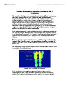
One of the most important differences in specific antigen recognition between the three types of molecules is the conformation of the antigen they recognize. One way to demonstrate this difference experimentally is to immunize an animal with a protein in a particular conformation and subsequently challenge the primed animal with the same protein in a different conformation. It can be shown that a B-Cell response only ensues if the tertiary structure of the protein used for immunization and challenge are the same. In contrast, a T-Cell response ensues even if the protein is in its native form during immunization and denatured during the challenge and vice versa. In addition, Ab determinants have been shown to be located on the surface of a native antigen, whereas TCR determinants may be buried within the core of the folded protein. The conclusion drawn from these experiments is that TCR and MHC molecules recognize linear epitopes on short peptides derived from the protein, whereas Ab molecules specifically recognize conformational determinants of the immunogen (which need not necessarily be protein). Ultimate proof for this model of specific antigen recognition came from elusion and analysis of the antigens bound by each of these molecules and X ray crystallography.
Diversity
The MHC exists as a finite number alleles and the peptide-binding region is germline-encoded. For this reason, MHC molecules must be promiscuous to present the enormous range of possible peptide antigens. Multiple lines of experimental evidence support this idea. Firstly, if transgenic T cells with specificity for a defined peptide-MHC complex are stimulated by APCs presenting that peptide, the response can be inhibited by addition of excess of structurally similar peptide. Secondly, transgenic expression of defined MHC alleles in insect cells (which do not normally have MHCs) showed that many different peptides can be eluted from these. Using experiments like these, it has been estimated that each allele can bind around 2% of all possible peptides.
In contrast, both TCR and Abs are highly specific. In the case of Abs, this specificity has been classically demonstrated by Landsteiner in the 1930s. It can be achieved because TCR and MHC genes undergo somatic rearrangement. This allows them to recognize antigens with a much higher specificity. It has been estimated that the naïve T-cell repertoire consists of 25-100 million distinct clones,, each of which recognizes only very specific determinants. One might argue that the B-cell repertoire is even larger for two reasons. On the one hand they do not undergo as rigorous negative selection in development and on the other hand, somatic hypermutation increases diversity of Ab specificities.
Affinity maturation & Avidity maturation
Another major difference in Ag recognition between the three molecules is their affinity. This can be measured directly by equilibrium dialysis.
MHC molecules have a Kd of 10-6 M. This does not change during an immune response.
In contrast, the Ag binding site of Abs has a Kd of around 10-7 M, which may increase during the course of the immune response to 10-11 M by affinity maturation. The mechanism of this is a process of somatic mutation. In addition, B-cells have the means to change the avidity of their antibodies by class-swiching.
T-cells lack the genetic mechanisms for somatic mutation of their TCR to increase affinity. However, Slifka and Whitton found that functional avidity of the T-cell response increased from 3.5 * 10-8 M 4 days after immunization to 5 * 10-10 M by day 8. Invariant MHC-tetramer staining indicated that the affinity of the TCR had not increased. As a mechanism for this avidity maturation of TCRs, topological changes of the composition of T-cell rafts has been put forward.
Signalling
Binding is not enough for specific antigen recognition. Crucially, recognition involves signal transduction across the plasma membrane.
It is not clear whether the MHC transduces a signal back to the antigen-presenting cell and analysis of the intracellular domain has not yielded any evidence for a role in signalling. However, the type of MHC carries information concerning the compartment from where the antigen was sampled.
Binding of the TCR to a peptide/MHC complex on the other hand results in transduction of a signal to the T-cell. Several lines of evidence support this idea and indicate how this might be achieved. The TCR forms a complex with trimeric CD3. Anti-CD3 antibodies may activate T-Cells in a similar manner to binding of the MHC/peptide complex to the TCR, if the T-cell line does not depend on co-stimulation. In addition, chimeric molecules have been engineered where the extracellular and transmembrane domains of the IL-2 Receptor were engineered on the cytoplasmic domain of CD3. Binding of IL-2 to these chimeric receptors had identical effects to MHC/peptide binding.
Similarly, membrane-bound antibodies are form a complex with Igα and Igβ. These transmembrane proteins signal specific antigen recognition when the BCR is crosslinked by the binding of its antigen. Secreted antibodies on the other hand can activate immune cells with their Fc receptors. In addition, they may activate the classical pathway (in the case of IgA also the alternative pathway) of complement, which serves to amplify the danger signal.
Conclusion
MHC, TCR and Ab molecules achieve specific Ag-recognition very differently. Importantly, the knowledge about these different mechanisms has helped us devise rational therapy and improve differential diagnosis.
For instance, altered peptide ligands have been engineered to antagonize the function of T cells in autoimmune disease. This is a direct consequence of the model of specific antigen recognition by TCR outlined above, and has been achieved by changing the peptide residues of the peptide that make contact with the TCR.
In the case of pemphigus vulgaris, the model of specific Ag recognition by the MHC has been able to explain the pathogenesis of an autoimmune disease. In HLA-DRB1*0402, the P4 pocket is formed by negatively charged amino acids. These can make a strong association with certain peptides processed from desmoglein, whose P4 residues carry a positive charge. This strong association leads to a T-cell response and autoimmune disease. In contrast, the wild-type HLA-DRB1 molecules do not have negatively charged residues at the P4 pocket. Screening for this particular mutation in the class II MHC has helped differential diagnosis and might lead to the ability to prevent the disease before it has become clinically apparent.
T Konrad Rajab – 1465 words
Arstila et al. „A direct estimate of the human alpha beta T cell receptor diversity”. Science 1999; 286:958
von Andrian et. al. „T cell Function and Migration” N. Engl. J. Med. 343: 1020
Silfka MK & Whitton JL, Nature Immunol. 2: 711 (2001)
Margulies DH, Nature Immunol. 2 : 669 (2001)