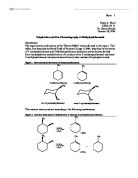
- Mandelin test
- Marquis test
- Scott test
- Erlich test
- Liebermann test
- Dille-Koppani test
- Duquenois-Levine test
- Microcrystalline tests
Various separation techniques could be used, such as high performance liquid chromatography & gas chromatography. Also analytical techniques such as mass spectrometry, infra red spectrometry & 13C NMR spectroscopy could be used before or after separation has been achieved.
Materials: The apparatus used in this experiment are listed below.
- Filter Paper
- Aspirin, Paracetamol & Caffeine samples
- Unknown Samples A,B,C &D
- Small TLC Chamber
-
Solvent 1= Ethyl acetate, Chloroform, 85% Formic acid (3:2:1)
-
Solvent 1= Ethyl acetate, Chloroform, 85% Formic acid (3:2:1)
-
Sample Solvent= 50% chloroform, 50% methanol
- Capillary Tubes
- TLC Plates
- U/V Lamp
Method: Student 1
Preparation of TLC solvent
The following solvents were provided:
Solvent 1 → Ethyl acetate, Chloroform, 85% Formic acid (3:2:1)
Solvent 2 → Ethyl acetate, Acetone, Butanol, 10% Ammonia (5:4:3:1)
Sample Solvent → 50% chloroform, 50% methanol
Solvent 1 was placed into the small TLC chamber to a depth of around 6mm & a watch glass was placed over the top to allow the vapour to fill the tank.
Preparation of samples
A micro spatula was used to put a small amount of each unknown sample (A,B,C & D) in to a cell in a test tray. Each of which was dissolved in a minimal volume of the sample solvent, this was also done with the aspirin sample to be used as a control.
Preparation of applicators
The applicators are prepared by holding capillary tubes at the tip of the blue flame provided by a Bunsen burner. When the capillary tube begins to glow red the two halves are quickly pulled apart. This produces a stretched & hence thinner end on the heated part of the tubes. The thinner circumference of the tubes allows for a more careful application of the samples & allows the samples to be collected using the tension.
Preparation of TLC plate
A pencil line is drawn 1 cm inside one edge of two TLC plates. This line must be drawn in pencil, if a pen was used the components of that pen would run as the samples do. On which three marks are made 1 cm apart labelled Aspirin A B on the first plate & Aspirin C D on the second. Great care is taken not to damage the delicate gel coating.
Application of samples
A sample of each ink is spotted onto the appropriate margin cross ensuring that each spot is no bigger than 2-3 mm across. The TLC plate is then carefully placed into the chromatography tank leaning one edge against the wall of the tank & the lid is replaced to allow the plate to develop.
Marking & recording Data
When the solvent front has travelled a sufficient distance of the plate it is removed & the solvent front immediately marked using a pencil. The plate is then allowed to dry before each spot that is visible under normal light is marked in the centre using a pencil. This is then repeated under UV light & the distance that the solvent front & each spot has travelled from the margin line is measured & the Rf value of each spot is calculated. It is important to calculate the Rf value to the centre of each spot as this is the most concentrated region of the spot.
Student 2 also does the above process using paracetamol instead of aspirin & student 3 does the above process with caffeine.
The whole of the above process is repeated using solvent 2.
Results:
Table 1 Rf value of each compound in solvent 1
Table 2 Rf value of each compound in solvent 2
From the above information the following can be concluded:
Unknown A contains the following compounds:
Unknown B contains the following compounds:
Unknown C contains the following compounds:
Unknown D contains the following compounds:
- Paracetamol
- Aspirin
- Caffeine
The following graphs will help to confirm this:
Graph 1: Solvent 1, Student 1 (Aspirin)
Graph 2: Solvent 1, Student 2 (Paracetamol)
Graph 3: Solvent 1, Student 3 (Caffeine)
Graph 4: Solvent 2, Student 1(Aspirin)
Graph 5: Solvent 2, Student 2 (Paracetamol)
Graph 6: Solvent 2, Student 3 (Caffeine)
Conclusion: Unknown Sample A: To look at student two & student three’s TLC results it is clear to see that it consists of two compounds. Unfortunately this is not apparent in student one’s findings, reasons for such may be due to human error. Fortunately in this case it appears that caffeine is not present in the mixture anyway. By looking at graphs 2, 3, 5 & 6 a clear comparison can be made although it is not quite as obvious in graph 3. The clear comparison on graph five justifies the statement that this mixture consists of the two compounds paracetamol & caffeine.
Unknown Sample B: We can see this mixture consists of two compounds, by comparing the Rf values on table one & on graph one & graph two there is a good comparison shown towards the control. Unfortunately the results found from solvent two did not show a sufficient comparison, in forensic practice the class of drug used would have determined the solvent used. The results from this experiment have led me to believe that solvent two is not as appropriate as solvent one for these particular drugs. Fortunately the results from using solvent one give good justification to the statement that unknown sample B mixture consists of the two compounds Paracetemol & aspirin.
Unknown Sample C: Initial conclusions can be taken by looking at the tabulated results that this is a mixture of two compounds (also noticeable from the fact that the TLC resulted in two spots). From the comparison of the controls & the tests from graphs one, three, for & six the constituents of this mixture become more apparent. Both solvent one & two showed a good comparison for evaluation & backed up the statement that unknown sample C contains the compounds caffeine & aspirin.
Unknown Sample D: It becomes immediately apparent that this mixture contains three components. In our situation we have been informed that the mixtures contain some or all of the following compounds:
- Caffeine
- Aspirin
- Paracetamol
So from this information we can immediately conclude that this unknown contains all three of the possible compounds. However in a real laboratory situation this would not be the case. Where we can make an immediate conclusion the forensic team would not have such a narrow row of possibilities. Hence further examination would be required. For this mixture comparison of all the above graphs is required, & clear comparison can be seen on all graphs with each control. So confidence can be shown in the statement that unknown sample D contains all three compounds (caffeine, aspirin & papacetamol).
General analysis of the TLC plates shows the constituents of each mixture. The further calculations of Rf values gave greater graphical information for comparison once tabulated & the graphs were made. The first thing to be noted from the plate should be that the colouring of each spot matches, only when this is noticed may we justify any calculation. Overall the experiment proved to be generally successful showing only small differences between the controls & the tests. The only large problem appeared to be the lack of agreement to student ones result which showed unknown sample A to only consist of one compound.
Evaluation: When we look at the TLC plates the spots are quite large, this is due to the plates being over loaded on application. Although the spots were only approximately 2-3mm in diameter as suggested the samples were too concentrated & hence we can see the results are less that perfect although suitable to give an answer to the aim. As a forensic application TLC (as a screening test) allows toxicologists to get a quick insight into the possibility of a sample containing a drug substance. Yet another advantage of using such tests is that they allow the analyst to analyse a large number of samples in a relatively short amount of time. One disadvantage is that such screening, & presumptive tests are only possible when there is a sufficient amount of specimen. In the forensic lab a positive tests from TLC is considered provisional & should be confirmed by instrumental analysis. The toxicologist follows a particular scheme demonstrated below in figure one:
The instrumental methods include those mentioned earlier, for example:
Figure 2: HPLC Equipment
Figure 3: GC-MS equipment
Such equipment would be used to confirm the identity of the drugs after initial screening has been completed (providing there is sufficient sample).
References: Table 1 Rf value of each compound in solvent 1
Table 2 Rf value of each compound in solvent 2
Graph 1 Solvent 1, Student 1 (Aspirin)
Graph 2 Solvent 1, Student 2 (Paracetamol)
Graph 3 Solvent 1, Student 3 (Caffeine)
Graph 4 Solvent 2, Student 1(Aspirin)
Graph 5 Solvent 2, Student 2 (Paracetamol)
Graph 6 Solvent 2, Student 3 (Caffeine)
Figure 1 Routes taken by toxicologist
Figure 2 HPLC Equipment
Figure 3 GC-MS equipment