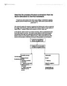
Peripheral nervous system
(Cranial and spinal nerves)
Somatic nervous Autonomic nervous system
System (under (under involuntary control)
Voluntary control) Sensory neurones/
- Sensory neurones/ visceral receptors
Afferent nerves Motor neurones/
- Motor neurones/ sympathetic and
Efferent nerves parasympathetic nerves
Davis. B, Bull. R, Roscoe. J, Roscoe. D; Physical Education and the study of sport; 1997 third edition; Mosby; London’ p 40
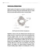
A motor neurone is started of as a cell body within the grey matter and its axon passes out of the ventral root of the spinal cord to innervate muscle cells. The muscle cell consists of three major parts that include a cell body containing the nucleus, mitochondria and other organelles, cellular extensions called dendrites and an axon. Dendrites look like fine branches with twigs extending out from a neurone. Following sensory stimulation a relay neurone transmits neural impulses to the dendrites of the motor neurone; they are specialised to receive electrical impulses and conduct these towards the cell body.
Axons are segmented tube-like extensions of a neurone. They arise from the thickened area of the cell body and emerge out of the ventral root of the spinal cord. The function of the axon is to transmit neural impulses away from the cell body towards muscle tissue or a gland. The surrounding myelin sheath acts as an insulator this will speed up the transmission of the impulse. Motor neurone typically divides up into several branches as it reaches the muscle bed. The branches then connect motor neurone to the muscle fibres by distinct structures known as motor end plates. Control by the brain is possible because of the ascending and descending fibres to the motor cortex.
Information is relayed from the brain to the muscle via a nerve impulse. A nerve impulse is an electrical current running the length of the nerve, starting at the brain and passing down the spinal column tot he relevant cell body. The cell bodies of individual motor neurones are located in various regions of the anterior horn of the spinal column. These collections of cell bodies are called motor neurone pools. The cell bodies are positioned in relation to the muscle they stimulate, for example the circumflex nerve, which stimulates the deltoid, is found in the fifth cervical vertebra.
The nerve impulse is passed along the axon of the motor neurone. The axon is covered in a myelin sheath. This sheath is mostly made up of fat to insulate the nerve, the sheath is not continuous. The node of Ranvier is place where there are gaps in the myelin sheath. The impulse is passed from one node of Ranvier to the next so that the impulse can travel faster, it slow down if it would need to travel the whole length of the axon. Saltatory conduction is the name of this nerve impulse propagation method. Also the thicker the myelin sheaths the faster the nerve impulse that can be conducted.
Once the impulse the end of the axon the nerve transmits the information to the muscle by releasing a chemical transmitter called acetylcholine, at the neuromuscular junction.
The nerve impulse is when information is relayed from the brain to the muscle via a nerve impulse. A nerve impulse is an electrical current running the length of the nerve, starting at the brain and passing down the spinal column to the relevant cell body. The cell bodies of individual motor neurones are located in various regions of the anterior horn of the spinal column. These cell bodies are referred to as motor neurone pools. The cell bodies are where the muscles are they stimulate for example the circumflex nerve.