Myofibrils contain protein filaments called ‘Actin’ and ‘Myosin’, actin and myosin move in opposite directions to cause muscular contraction, muscles shorten no more than about 60% of the resting length.
Parts of the microfibril: Actin and Myosin protein filaments make up much of the muscle fibre, Actin is the thin, light filament, and myosin is the thicker, darker filament. The arrangement of theses fibres produces darker and lighter areas; the dark areas of myosin are called ‘A bands’ and the light areas of actin are called the ‘I bands’. Actin filaments can also occur inside the A bands, during contraction, the actin filaments slide further into the A bands. The area in the middle of the A band that has no actin is called the ‘H zone’, myosin filaments grow thicker here and are called the ‘M line’, during contraction, this area and the I bands grow smaller due to the actin filaments. In the I band, the actin filaments are connected in a zig-zag arrangement.
Structure of Actin and Myosin Each of these are protein filaments, Myosin is the most abundant, it accounts for about two thirds of the muscle protein. The molecule is composed of two twisted protein strands; many of these molecules form a filament. The strands have globular parts called ‘cross-bridges’ which project outwards. Actin accounts for about a quarter of the muscle protein. The molecule is a globular structure composed of ADP molecules, these molecules serve as active sites for the cross bridges. Filament is formed form double twisted strand (helix) of actin molecules.
Other muscle proteins Tropomyosin and Troponin (each are associated with the actin filament)
Tropomyosin is a rod-shaped muscle protein that occupies the longitudinal grooves of the actin molecule. Troponen is a molecule on the tropomyosin and is called a tropomyosin / troponen complex.
How contractions occur Muscle at rest: Troponen/ tropomyosin complex is exposed to the myosin, no linkage can be formed between these two molecules. Contraction: High concentration of Calcium Ions becomes present; these Calcium Ions bind to the troponin. This moves the position of the complex and exposes the active sites on the actin to the myosin cross bridges, linkages can be formed which causes the shortening.
Ratchet theory: A linkages is formed between actin and myosin filament, the cross bridges on myosin bends, causing some shortening. This lines up another actin myosin site, so another linkage / shortening occurs, this causes the original linkage to break and it is now free to form another.
The Calcium comes from the SR, which contains a high level of Calcium Ions. The action potential reaches the SR and the membrane becomes more permeable to Calcium Ions.
Muscle contraction needs stimulation from a specific neurotransmitter (acetylcholine), which is produced in the cytoplasm of motor neurones. It is then shipped in vesicles to the motor nerve fibre where it remains; the nerve impulse then reaches the end of the axon. Acetylcholine is released into the gap between the axon and the motor nerve plate and binds with receptor molecules in the sarcolemma; this binding causes the action potential. Stimulus travels in all directions over the surface of the sarcolemma until it reaches deep into the muscle and finally to the SR.
Relaxation: An active transport system begins to work, this rapidly moves the calcium ions back into the SR, at this point, and calcium is moving both into and out of the SR. Also, ‘cholinesterase’ begins the rapid decomposition of acetylcholine, when the acetylcholine gone, the SR is no longer permeable to calcium. The linkages between actin and myosin break and troponen is exposed, therefore muscular contraction is inhibited.
Muscular contraction review: Mentally, when we want to contract a muscle, the nerve cell that controls it sends an impulse towards the muscle. When the impulse reaches the neuromuscular junction (synaptic gap) ‘Acetylcholine’ is released. Acetylcholine binds with the receptor molecules in the cell, which causes and action potential (AP) to be generated in the muscle. The AP reaches deep into the muscle to the sarcoplasmic reticulum (SR) and the AP causes the SR to become more permeable to Calcium (Ca). Calcium leaves the SR and binds with Troponin (located on the Tropomyosin); this causes the tropomyosin-troponen complex to invert itself. This exposes the active sites, which bind to the myosin cross bridges. The presence of ATP causes bending at this connection, resulting in muscular contraction.
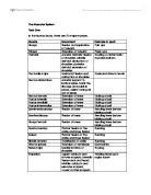
There are 3 main types of contraction: Static contraction – isometric muscle contraction. Concentric contractions – isotonic and Isokinetic muscle contraction and eccentric contraction.
Isometric muscle contraction is a static contraction, which occurs when a muscle develops tension but remains at the same length.
Isotonic muscle contraction is a dynamic contraction in which the sports performer controls the speed of contraction.
Isokinetic muscle contraction is a dynamic contraction, which occurs when the rate of movement of origin towards insertion of a muscle is constantly maintained throughout a specific range of motion even though maximal force may be exerted.
Eccentric muscle contraction occurs when a muscle is activated and force produced but the muscle lengthens.