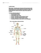
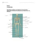
Pelvic bones
The pelvis varies in shape according to sex. Overall, the structure has a similar appearance in both sexes but takes a shallower and wider form in females to allow for the specialised function of childbearing. Arranged in a ring, the fused pelvic bones provide a strong foundation for the upper body and protection for parts of the reproductive systems.
Bones of the skull
Two separate sets of bones from the intricate structure of the skull. The eight bones enclosing and protecting the brain are called the cranial vault. Another 14 bones make up the skeleton of the face. In adults, all of the lower jaw, (mandible) are locked together by joints known as sutures. Theses seams are visible on the surface of the skull as lines between the bones.
Viewed from the front, the most prominent skull bones are the frontal bone, which forms the forehead, the zygomatic bones, which give shape to the cheeks, and the upper and lower jaw bones. The back and sides of the cranial vault largely comprises the occipital and parietal bones. In the middle ear there ate three tiny bones of the type known as ossicles, which are not technically part of the skull. They conduct sound waves to the ear drum to the inner ear.
Structure of the spine
The spine is made up of 33 ring liked bones called vertebrae they are linked by a series of mobile joints. Sandwiched between the vertebrae are springy, shock absorbing disks with a tough outer layer of cartilage. There are three main types of vertebrae: cervical in the neck, thoracic in the upper back, and lumbar in the lower back. Two regions at the base of the spine, the wedge shaped sacrum and the tail likes coccyx, both consists of several fused vertebrae.
Coccyx – the four tale bones of the spine are much smaller than the other vertebrae.
Sacrum – the five vertebrae are fused
Spinal nerve – connected to the spinal cord are 31 pairs of nerves that emerge through gaps between the vertebrae and travel out to body tissues and organs
Vertebral body – these bony disks become larger towards the base of the spine to support greater weight
Vertebral processes – these bony knobs extend from the back of each vertebra. Three processes serve as anchor points for muscles; the other four form the linking fact joints between adjacent vertebrae.
Spinal cord – this vital cable of nerve tissue, which relays messages between the brain and the different parts of the body, is protected by the 33 vertebrae of the spinal column
Intervertebral disks – composed of though, flexible cartilage with a jelly like core, these disks protect the vertebrae from pressure.
Facet joint – this linkage point between the vertebrae is formed by the round ended process of one bone fitting into a matching hollow in the process of the bone above
Atlas - this is the top most cervical vertebra to which the skull is attached.
Curves of the spine
A healthy spine has 4 curves that help to make it resilient and maintain balance. The cervical and lumber section curve forwards, while the thoracic and sacral sections curve back wards. Abnormal curves may be due to poor posture, congenital defect, or bone disease.
Reagons of the spine
Each section of the spine is adapted to a particular function. The cervical vertebrae support the head and the neck, the thoracic vertebrae anchor to the ribs, and the strong, weight baring regions towards the bottom of the spine provide a sable centre of gravity during movement. Although their shapes vary vertebrae typically comprise a bony disk called the body projections called processes to which muscles attach.
Cervical vertebra – a typical cervical vertebra has two wing shaped side processes. A whole through each process allows arteries to pass through and carry blood to the brain.
Thoracic vertebra – the thoracic vertebrae form part of the protective rib cage in the thorax. Each of these vertebrae has small hollows in the processes and body into which a pair of ribs fits.
Lumber vertebra – the larger body of a lumber vertebra reflects is role in supporting a major part of the body’s weight. The articular processes facilitate movement.
Movement of spinal joints
The spine is constructed strongly to hold the head and body upright, but it is also flexible enough to allow the upper body to bend and twist. The cartilage disks between the vertebrae can withstand enormous forces, which can be as much as several hundred kilograms per square centimetre during strenuous movements. Strong ligaments are muscles around the spine stabilise the vertebrae to help control movement.
Ligament – stabilizes vertebrae and holds them in alignment during movement.
Facet joint – helps to determine the degree of movement between vertebrae.
Intervertabral disk – absorbs forces directed through its axis and acts like a ball bearing during bending and twisting.
Spinal joints - individual spinal joints do not have a wide range of movement, but working together they give the spine great flexibility, letting it arch backwards, twist around, or curve forwards.
Flexibility – the body is capable of bending further forwards that backwards; this is due to the shape of the vertebrae. The top 7 vertebrae which make up the cervical spine are the most flexible.
Bone structure
Bone is made up of specialised cells in a matrix composed mainly of protein fibres, water, and minerals. In the centre of the long bone is the medullary canal, which contains bone marrow and blood vessels. Around the marrow are layers of spongy (cancellous) bone, which also contains marrow, and hard (cortical) bone. A membrane, the periosteum, covers the bone surface.
Periosteum – this thin, fibrous membrane covers the entire surface of bones except inside the joints. Its blood vessels supply nutrients, and its nerves signal pain.
Medullary canal – this canal contains red bone marrow, which produced blood cells, and yellow marrow, which is mostly fat tissue
Long bone – at each end of long bones of the limbs there is an area known as the epiphysis. In childhood, the epiphyses are made mostly of cartilage, which hardens to become spongy (cancellous) bone in mature adult. The central shaft of the long bone is called the diaphysis.
Osteon – osteons, which are also called haversian systems, are the building blocks that make up hard (cortical) bone. They are rode shaped units with a central canal that is surrounded by concentric layers of bone tissue called lamellae.
Lamellae – in each layer, fibres of collagen, a type of protien, face different directions for added strength.
Osteocyte – this basic bone cell is located in the gaps (lacunae) in the bone matrix.
Bone growth
Between the shafts (daiphyses) and the ends (epiphyses) of the long bones is a growth area known as the epiphyseal plate. Cartilage cells, known as chondrocytes, proliferate here and form columns that push older cells towards the middle of the bone shaft. The cartilage cells enlarge and eventually die; leaving a space that is filled by new bone cells. Bone growth continues until about the age of 17.
Bone repair
Bone is a living tissue that is continually broken down and rebuilt throughout a person’s life. If it is fractured, bone is able to regrow, and eventually the line of fracture is bridged with new tissue. The repair mechanism is activated rapidly after injury, although the laying down of new bone may take weeks to complete. Once mended, a broken bone may take some months to regain its full strength.
Joint structure
A joint, or articulation, is where two bones meet. Joints are classified by their structure or by the way they move. Most joints in the body are synovial joints. These are versatile, lubricated joints, such as the knee, in which the surfaces in contact slide over each other easily. Articular cartilage covers the bone ends, ligaments provide stability, and fibrous capsule encloses the joint. Surrounding muscles produce movement.
Fixed and semi-movable joints
Not all joints are freely movable. After growth is complete, the bones of the skull become fixed together by fibrous tissue, forming immovable suture joints, in the lower leg, the tibia and fibula are stabilized by ligaments that allow only a small amount of movement.
Types of synovial joint
Ina synovial joint, the shape of articular cartilage surfaces and the way they fit together determine the range and direction of the joints movement. Hinge and pivort joints move only in one plane for example from side to side or up and down, while ellipsoidal joints are able to move in two planes at right angles to each other. most joints in the body can move in more than two planes, which allows for a wide range of movements.