The Structure of Prokaryotic and Eukaryotic cells.
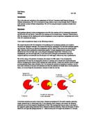
The Structure of Prokaryotic and Eukaryotic cells The prokaryotic cell developed first, pro meaning before and karyon meaning nucleus. The evolution of prokaryotic cells preceded that of eukaryotic cells by 2 billion years. This group of cells are now known as bacteria and archae. This simple cell has evolved into the eukaryotic (true nucleus) cells, a more complex cell, generally found in higher organisms such as plants and animals. The fundamental difference between the two cells is the presence of a nucleus in the eukaryotic cell. Though the prokaryotic cell contains genetic information, it is suspended freely in the cytoplasm. The eukaryotic cell is also considerably larger than the prokaryotic, it possesses an endoplasmic reticulum along with many other complex organelles and synthesises larger ribosomes. On the other hand all prokaryotic cells have cell walls, where as it is generally only the eukaryotic cells of plants that possess cell walls. In this essay, the similarities and differences between these two cells will be discussed along with exploring which cell is more primitive and why. The Prokaryotic Cell The Eukaryotic Cell Scientists believe that the eukaryotic cell originated from several different types of prokaryotic cell, hence the similarities between the two cell types. The contents of both cells are suspended in the cytoplasm, due to the merging of

Changes in Temperature affecting Amylase and Starch
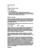
Valentina Zunarelli IB Biology Practical Changes in Temperature affecting Amylase and Starch Aim Testing whether a change in temperature will affect the way amylase, an enzyme, acts upon starch (breaking it down), and finding amylase's optimum temperature. Apparatus · Bunsen Burner · 10ml measuring cylinder · Thermometer · Stop clock · Iodine solution · Starch · Amylase · Spotting tile · Pipettes · Two test tubes · Tongs Method Firstly, fill the spotting tile indents with iodine (about 5 drops) using a pipette. Then, in two test tubes separately, heat 5ml of amylase solution and 10 ml of starch solution starch. Keep heating over a bunsen burner holding the test tubes with tongs until the temperature of the solutions is 30?C, use a thermometer to be accurate. Once the temperature has been reached mix the two solutions together by pouring the starch solution in with the amylase (swirl the test tube to make sure the solutions mix). As soon as this is done, start a stop watch and every twenty seconds, using a pipette, take some of the mixture and add two drops of it into a different indent on the spotting tile with the iodine each time. The iodine should change colour; this change should be recorded. Next, the experiment should be repeated but instead of heating the solutions to 30?C, they should be heated to 40? then 50?, 60?, 70?C and lastly 80?C,
An experiment to investigate how temperature affects the permeability of beetroot cell membranes
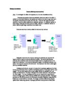
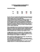
An experiment to investigate how temperature affects the Permeability of beetroot cell membranes Interpretation of Results: 0°C 20°C 30°C 40°C 55°C 0.24 0.28 0.75 0.79 0.22 0.26 0.35 0.41 0.81 From 0°C to 30°C there is a gradual increase of absorbency which shows that as the temperature increases it is denaturing the cell wall and cell membrane and allowing the beetroot pigment to leek out into the distilled water. As soon as the sample of beetroot is placed into the distilled water diffusion occurs naturally, which is the net movement of molecules from a region of high concentration (beetroot sample) to a region of low concentration (distilled water). From 40°C upwards there is a sudden increase of absorbency, this is because the optimum temperature of which enzymes and cells can work at is 37°C and as soon as you go above this temperature it will cause the enzymes and cells to denature and cease from working as effectively. Even if you then lower the temperature again the cell will not regain its original shape. Within the phospholipid bi-layer there are proteins, and these proteins are made up of polypeptide chains which are joined together by hydrogen, hydrophobic and peptide bonds. Once the temperature has increased above 40°C the molecules vibrate so energetically that these bonds break easily and therefore creating holes within the cell

Investigating the effect of substrate concentration on reaction between Hydrogen Peroxide (H2O2) and the enzyme Catalase.
Investigating the effect of substrate concentration on reaction between Hydrogen Peroxide (H2O2) and the enzyme Catalase AIM Catalase is an enzyme found in Yeast, and when yeast reacts with H2O2, a reaction occurs, with H2O2 acting as the substrate, and Catalase being the complementary enzyme. In this experiment, I will analyse, interpret, conclude and evaluate the effects of reacting different concentrations of H2O2 with the enzyme Catalase and hence, use it to understand the effects on the rate of the reaction. My main aim is to understand how enzymes work under varied conditions. Yeast is not a chemical and has no chemical formula. Try looking at yeast as a single cell, similar to any single cell in your body. Those cells are made up of numerous types and classifications of chemicals. Some are very simple, like water. Others are extremely complex, like proteins. As the enzyme Catalase is not available in pure form, I will use yeast as the source of enzyme. There are other alternatives e.g. potato tubes which also contain catalase, but using yeast would give me greater accuracy because the amount of Catalase in each potato tube would be different. BACKGROUND INFORMATION Enzymes are biological catalysts, which means that they speed up biological reactions without getting used up themselves. They also provide a control mechanism for reactions, as the amount of

Investigating the Effect of Temperature on Rate of Respiration.
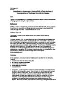
* Biology A2 Coursework Name: Luke Meredith Form:13 CJN PLANNING Title: Investigating the Effect of Temperature on Rate of Respiration Aim: I am to investigate and take readings of oxygen consumption, on an organism at several temperatures including replicate readings to give a mean value. At each temperature I will leave the invertebrates for about 10 minutes for the rate of respiration to reach equilibrium. Diagram Experiment 1 Experiment 2 Partial 2b . Complete 4c. Apparatus List: Test tubes x2 Invertebrates (maggots) Respirometer Data Harvesting software on computer and probe Gauze platform clamp stands cm syringe Glass beads Sodium hydroxide . Preliminary Work Conducted The first diagram shows two test tubes linked to respirometer. One test tube contains invertebrates (in this situation, maggots) with sodium hydroxide underneath separated by a gauze platform. The other contains glass beads and also sodium hydroxide as shown on the diagram. The glass beads act as inert material to the volume of the invertebrates. Using this method I could measure the rate of respiration at different temperatures, of these invertebrates, by changing the temperature of test tube one and then take readings from the respirometer, from the U-tube containing manometer fluid. What should happen is the invertebrates will respire and the carbon dioxide
Structure and Function of Red Blood Cells and White Blood Cells
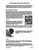
Structure and Function of Red Blood Cells and White Blood Cells Bloods importance to human life has been recognised since pre-history, acquiring mystic and religious significance through the ages. Biologically, blood is a liquid tissue, transporting materials and protecting us against disease. Suspended in the watery plasma are red and white blood cells, serving different functions, but both of vital importance to our bodies. Red Blood Cells (RBC's or erythrocytes) RBC's are responsible for carrying oxygen (O2) and removing carbon dioxide (CO2) from about 30 trillion cells in the human body. In normal blood RBC's account for about 45% of the total volume. On average, we have about 5 million red cells per cubic millimetre of blood. Produced in the red marrow of bones, RBC's arise from a single type of cell, called a stem cell. During formation the nucleus is lost and organelles degraded, allowing more internal space to be filled with haemoglobin, the O2 carrying protein abundant in red cells. Without a nucleus RBC's can never divide. Lacking in organelles, they can only survive for about 120 days, before being 'eaten' by white cells. Some 3 million RBC's die and are scavenged by the liver every second. Structure RBC's are biconcave shaped discs (as seen in this electron micrograph), about 7.5µm diameter. The shape gives an increased surface area, allowing the
An experiment to demonstrate bacterial staining.
Liverpool John Moores University in Association with St Helens College Franchise Degree of B.Sc. (Hons) Applied Biology Basic Microbiology An Experiment to Demonstrate Bacterial Staining Assignment 1 Introduction If bacterial colonies are measured to be between 1mm to more than 1cm, then it is obvious that the individual microbe is a lot smaller. In this experiment to observe the microbes more thoroughly, techniques that were founded, tried and tested many years ago such as various staining techniques, were used to observe the individual microbes in this experiment for their characteristics, and with the aid of a light microscope this was possible. The purpose of staining in microscopy is to: - Add contrast to the image Identify chemical components of interest Locate particular tissues, cells or organelles. The three techniques carried out in this experiment; namely Gram Staining, Endospore Staining and the Hanging Drop; shown characteristics like size, form, elevation, colour and motility. Even under light microscopy (conditions better than the human eye), the bacterium portrayed clearer once it had been stained. Microbial characteristics have in the past proved to be of importance to mankind, having the knowledge regarding bacteria created many medicinal uses, such as the production of antibiotics and led to great advances in finding cures for deadly diseases,
Experiment to Investigate a Factor which Affects the Rate of Decomposition of Hydrogen Peroxide by Catalase.
Phil Cooper L6 0.11.01. Experiment to Investigate a Factor which Affects the Rate of Decomposition of Hydrogen Peroxide by Catalase Aim The aim of this investigation is to investigate a factor which affects the rate of decomposition of hydrogen peroxide using the enzyme catalase. Background Catalase is present in nearly all the peroxisomes of nearly all aerobic cells, serving to protect the cell from the toxic effects of hydrogen peroxide by catalysing its decomposition into molecular oxygen and water. The overall reaction for this is: 2 H2O2 2 H20 + O2 The enzyme is among the most efficient known, with rates up 200,000 catalytic events/second. 'Hydrogen peroxide, H2O2, is a colourless, syrupy liquid that is a strong oxidising agent and, in water solution, a weak acid. It is miscible with cold water and is soluble in alcohol and ether. Although pure hydrogen peroxide is fairly stable, it decomposes into water and oxygen when heated above about 80°C; it also decomposes in the presence of numerous catalysts, e.g., most metals, acids, or oxidisable organic materials. A small amount of stabiliser, usually acetanilide, is often added to it. Hydrogen peroxide has many uses. It is available for household use as a 3% (by weight) water solution; it is used as a mild bleaching agent and medicinally as an antiseptic. The 3% solution is sometimes called ten volume
The purpose of this experiment was to investigate the interaction of different dyes to mammalian cells and tissues, to thereby determine their effectiveness in comparison with each other.
INTRODUCTION The purpose of this experiment was to investigate the interaction of different dyes to mammalian cells and tissues, to thereby determine their effectiveness in comparison with each other. Although the purpose of histological research is to try to study tissues in their most natural state this is impractical as most tissues look fairly similar without some kind of preparation. For this reason staining has become one of the most important procedures in the diagnosis of pathological conditions, as staining cell samples allows in-depth analysis of almost every aspect of the cell. For this experiment two of the most widely used dyes were used, those being methylene blue, a cationic stain, and haemotoxylin and eosin, a complimentary stain which is made of a cationic and a anionic mixture. The purpose being to decide which of the two stains is the most suitable for the use in analysing rat kidney tissue samples. Each dye will be used in various mixtures and pH values (as per the method) to see which dye produces the best results, i.e. the clearest and more distinguishable cells and organelles. When soaked in dye the cells and organelles change colour this is due to a process of ion exchanges. In each cell the chromagens (the staining part of the cell) exchange the cations in the tissue, such as Na+ and H+ with larger ions from the stain. Tissue- Na+ + Dye+

The power output of a solar cell is proportional to the sine of the angle between the incident light and the face of the solar cell.
Jack Webdale 01/05/2007 Page 1 Proving the lens formula Hypothesis The power output of a solar cell is proportional to the sine of the angle between the incident light and the face of the solar cell. Apparatus I will use the following equipment during me experiment. A Solar Cell - A device used to convert light (photons) into Electric (Voltaic) that can be used to power circuits. A Metre Rule - Will be used to measure the distance between the light source and the solar cell. Will maintain the distance as accurately as possible. It has millimetre units, although large and hard to take a precise measurement in mm. Two Digital MultiMeters - These will serve the purpose of an Ammeter and Voltmeter. Using the different settings they can be used to give 2.d.p readings. A Resistance Box - A device used to serve as a fixed resister, but can serve as many sizes. For this experiment I will set it to 5ohms. A Light source (Mains Voltage) - small Lamp, 40-watt bulb. Used to emit light to give energy to the solar cell A Protractor -Used to measure the angle being used for each reading when turning the Solar cell. Connecting Wires - used to complete the circuit and connect the components. Most of the apparatus I will use are self-explanatory. I am satisfied that the experiment will be conducted as accurately as possible, although I can see it will be difficult to keep the