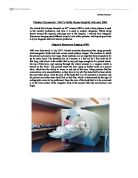
Magnetic Resonance Imaging.
Magnetic Resonance Imaging
In 1944, Isidor Isaac Rabi was awarded the Nobel Prize for Physics for his resonance method for recording the magnetic properties of atomic nuclei. This method was based on measuring the spin of the protons in the atom's core, a phenomenon known as nuclear magnetic moments. From Rabi's work, Paul C. Lauterbur and Peter Mansfield were able to research into magnetic resonance imaging (also known as nuclear magnetic resonance, NMR) and were awarded the Nobel Prize for Medicine in 2003.
Lauterbur, a professor and director of the Biomedical Magnetic Resonance Laboratory at the University of Illinois, realised that it was to possible to create an 'internal picture' of an object by NMR and had his ideas witnessed by a colleague. These ideas were based on the use of a magnetic field gradient - a magnetic field that varies through space.
Mansfield, a professor of physics at the University of Nottingham had no knowledge of Lauterbur's work and had an idea of how he might get an NMR picture of a crystal, similar to an X-ray signal crystal structure. With continual pioneering work with his colleagues, he was able to produce the first picture from a live human subject in 1976 with true anatomical detail. He continued to be a pioneer in the field, developing better imaging methods for larger body parts and also for imaging well past the sub-cellular level, all using the idea of NMR.
How does MRI work?
To investigate this, I intend to give an account on the basic physics of MRI and then explain the significance of the phenomenon to today's society. The areas I will research and address are:
* The basic idea of MRI
* The gyromagnetic ratio
* Speed of precession in a magnetic field
* Larmor Frequency
* Relaxation times
* Gradient Fields
* Production of a magnetic resonance image
* The major advantages and disadvantages of the system.
Basic idea
Hydrogen nuclei are subjected to a pulse of radio waves, which causes them to briefly emit low-intensity radio waves. These are detected by the MRI scanner, which measures the signal as the patient is scanned by a changing magnetic field. The signal is processed, producing an image of where the hydrogen atoms in water molecules and lipids are located.
Nuclei used in NMR contain an odd number of protons or neutrons and so possess intrinsic spin. (Spin is an inherently quantum property. In the context of this document, it refers to the turning motion of a proton). Hydrogen nuclei also possess a magnetic moment M (a magnetic field created by the moving positively charged protons) that has its origins in circulating currents. This means that there is always an angular momentum J associated with it. The vector quantities M and J are related by the equation:
M = ?J
Where M = magnetic moment
? = The gyromagnetic ratio
J = angular momentum
Angular momentum is the momentum of a body, which undertakes rotational motion. Momentum, P itself is a vector quantity that determines the potential force that an object can impart to another object by collision. It is calculated by mass x velocity. Since angular momentum involves rotational motion, it can be calculated by:
Radius of circular motion x momentum
Since momentum = mv,
Angular momentum = mvr
The gyromagnetic ratio is a nuclei-specific constant. For the normal isotope of hydrogen, with one proton, one electron and no neutrons it is 42.6MHz/T.
The equation means that a system must possess a magnetic moment, angular momentum and experience torques for it to exhibit a magnetic resonance. Torques are turning forces causing rotational motion.
Precession
When nuclei are in the presence of an external magnetic field they experience torques trying to align them with the applied field. Since they are spinning, the resultant movement is a precession. In other words, the nuclei will rotate around the axis of the magnetic field. If the protons in the nucleus can be made to precess about the external field lines in phase with each other, then their transverse field components will add up to give a small net transverse field that rotates about the external field axis at the Larmor frequency. The Larmor frequency is the resonant frequency of the precession of the protons.(Resonance normally refers to a vibration or oscillation only occuring at a certain frequency. In this case, it refers to the protons absorbing energy only at the Larmor frequency).The field can be detected and produce the information from which the image is generated in a MRI scanner. This will be explained later in the production of a magnetic resonance image section.
In order to achieve the protons precessing in phase with each other, they must be made to absorb radio-frequency radiation of the same frequency as the Larmor Frequency of the precession. Protons which absorb this energy will flip from a low-energy state to a high-energy state with their axes anti-parallel to the external field lines.
(a) Shows random alignment of hydrogen atoms when there is no magnetic field applied.
(b) Shows that when a strong magnetic field Bo is applied, the protons, having their magnetic moments, align themselves with the external magnetic field. They precess along the field lines of the magnetic field. Precession is shown more clearly below:
[Source: http://www.cs.sfu.ca/~stella/papers/blairthesis/main/node11.html]
A spinning proton Precession
Z represents the direction of the direction of the external magnetic field as shown by B0. X and Y are the respective axes at right angles to the magnetic field.
The red arrows are the protons which are precessing around Z. They are in their low-energy state, parallel to Z.
[Source: http://www.erads.com/mrimod.htm]
The majority of protons will align parallel to the external magnetic field since this is when they are in a low-energy state. Some however, as shown in the diagram below will align anti-parallel in the high-energy state. These will cancel out the protons pointing upwards. The remaining protons in the low-energy state which have not been cancelled, add up to produce a magnetic vector in effect, in the direction of the B0 field. This is known as longitudinal magnetization.
[Source: "MRI made easy" by Prof. Dr. Hans H. Schild page 12, published by Nationales Druckhaus Berlin 1990]
Z is the uniform B0 field as before, and X and Y are again the respective axes at right angles to the field. The red arrows are the protons, and the elliptical white arrows show that the protons are not static but are precessing about the magnetic field line.
Larmor Equation
The Larmor frequency is proportional to the strength of the external magnetic field:
?0 = ??0
Where ?0 = Larmor frequency
B0 = external magnetic field
? = gyromagnetic ratio
This equation demonstrates that the stronger the external magnetic ...
This is a preview of the whole essay
Z is the uniform B0 field as before, and X and Y are again the respective axes at right angles to the field. The red arrows are the protons, and the elliptical white arrows show that the protons are not static but are precessing about the magnetic field line.
Larmor Equation
The Larmor frequency is proportional to the strength of the external magnetic field:
?0 = ??0
Where ?0 = Larmor frequency
B0 = external magnetic field
? = gyromagnetic ratio
This equation demonstrates that the stronger the external magnetic field, the greater the Larmor frequency. A greater magnetic field would result in a greater torque experienced by the nuclei and they would precess at a faster rate. The value for the Larmor frequency calculated using this equation is important as the radio-frequency pulse will need to have the same frequency as the Larmor frequency of the protons in order for them to absorb energy and resonate. (A radio-frequency pulse is a short burst of an electromagnetic wave which has its frequency in the radio wave region of the spectrum).
Relaxation times
When the Radio-frequency (RF) pulse is sent at the Larmor frequency, more protons flip to the high-energy state, anti-parallel to the external field as mentioned earlier. The reason why this pulse is required is because the protons left in the low-energy state will produce longitudinal magnetization which cannot be measured directly as it is in the same direction, parallel to the B0 field. Thus the pulse not only causes the protons to flip into their high-energy state but causes them to precess in synch, in phase with each other producing a transversal magnetization as their vectors now add up in the direction transverse to the external magnetic field.
[source: "MRI made easy" by Prof. Dr. Hans H. Schild page 21, published by Nationales Druckhaus Berlin 1990]
This diagram shows that once the pulse is sent, more protons precess anti-parallel to the B0 field and in phase, resulting in less longitudinal magnetization (in the upwards direction) and more transversal magnetization (towards the right). These components are shown as vectors. The vectors represent forces of a certain direction.
Once the RF pulse is removed, the protons return back to their low-energy state and the longitudinal magnetization increases. This is known as longitudinal relaxation and is described by a time constant T1. The transversal magnetization will decrease, as the protons are no longer forced to remain in phase and this is known as transversal relaxation, described by a time constant T2. Transversal relaxation also occurs as the field of the MR magnet is not totally uniform, not totally homogeneous and so slight variations will cause different precession frequencies of the protons as shown by the Larmor equation. This will result in them precessing out of phase.
The T1 and T2 time constant can be shown graphically as below. Magnetization (%) is plotted against time (ms).
T1 Relaxation curve
[source: "MRI made easy" by Prof. Dr. Hans H. Schild page 27, published by Nationales Druckhaus Berlin 1990]
T2 Relaxation curve
[source: "MRI made easy" by Prof. Dr. Hans H. Schild page 30, published by Nationales Druckhaus Berlin 1990]
The T1 recovery curve shows a steep initial increase of longitudinal magnetization where the relationship is directly proportional but it then increases at a slower rate and levels off when maximum longitudinal magnetization is reached.
The T2 decay curve shows a steep initial decrease of transversal magnetization but as time goes on, the magnetization decreases at a slower rate until minimum transversal magnetization is reached.
Both relaxation times are not however defined as the times when relaxation is completed. Instead T1 is defined as the time when about 63% of the longitudinal magnetization is reached and T2 is defined as the time when transversal magnetization decreased to 37% of the original value. These percentages are derived from mathematical equations describing signal intensity (63% = 1-1/e; 37% = 1/e) but further detail into this is not necessary.
The transversal component of the net magnetization is constantly moving, constantly changing and so induces an electrical current into the receiver coil as described by Faraday's law which is to be explained. This is the signal used in MRI. The rate of change of transversal magnetization over time is determined by the relaxation times and because different tissues have different relaxation times, the rate of change of the transversal magnetization will be different and the MR signal obtained will be of a
different intensity. (as differing amounts of currents are induced).
Where ? is the induced emf or voltage
N is the number of turns on the receiver coil
? is the magnetic flux
t is the time over which the magnetic flux changes
Faraday's law shows that the emf induced is proportional to the rate of change of magnetic flux (d?/ dt) multiplied by the number of turns on the coil, N. emf (electromotive force) can be thought of as the amount of energy a source can produce per unit charge, coulomb. In this case, it can be used as meaning the same as voltage. Magnetic flux can be thought of as the lines of force surrounding a permanent magnet. In MRI it is the magnetic moments of the protons, which add up to produce the transversal magnetization vector.
Since current is proportional to voltage; I = V/R a changing magnetic flux or changing magnetic moment will induce a current, the signal in the receiver coil.
Once the transversal magnetization has disappeared after the RF pulse has been applied, no signal will be present (no current will be induced) as longitudinal magnetization is completely restored. This means that another radio-frequency pulse must be applied for the signal to be produced again. Initially a 90º pulse can be sent to tilt the net magnetization 90º in the direction transverse to the external magnetic field allowing a signal to be produced. After a certain time when most of the transversal magnetization has disappeared, another 90º pulse can be sent causing the same effect. The time between these pulses is known as the time to repeat, TR and is important in distinguishing between tissues of different types, hence increasing the resolution of the image (the resolution being the smallest distance between 2 objects which can be easily distinguishable).
The strength of the signal obtained after sending the second 90º pulse, depends on the amount of longitudinal magnetization that we start with as this is tilted to become the transversal magnetization. If a relatively long time is elapsed between sending the second RF pulse, then the longitudinal magnetization will have recovered totally and the signal after the second pulse will thus be the same as the one after the first pulse.
Since different tissues have different longitudinal relaxation times, T1 values, using a short TR would mean that tissues with a short T1 would have more longitudinal magnetization than those with a longer T1 by this time. This results in more longitudinal magnetization being tilted by the next 90º RF pulse and a stronger signal produced. This difference in signal intensity allows different tissues to be differentiated between.
Images produced by using a short TR (typically less than 0.5 seconds) to make the differences in T1 between tissues pronounced, are known as T1-weighted images. T1 is not the only parameter which signal intensity depends on. When a long time has elapsed between one pulse and the next (long TR, more than 1.5 seconds), there may still be a difference in the intensities of the signals produced. This is mainly due to a difference in the proton density of the tissues. More protons in a given area in a tissue would result in more protons precessing in phase when an RF pulse is sent, and more magnetic moments of the protons will add up to produce a greater transversal magnetization vector. The consequence would be a greater rate of change of the magnetic flux as d?/dt in faraday's law as ? is larger and more current will be induced, hence a stronger signal produced. Images produced based on the density of protons are known as proton density-weighted images.
The transversal relaxation time, T2 is yet another parameter which signal intensity depends on. The pulse sequence used to produce T2-weighted images is different however. A 90º RF pulse is used first to tilt the longitudinal magnetization, producing transversal magnetization as before, and is then switched off. The protons then lose phase coherence as they have different precession frequencies and the transversal magnetization decreases and thus the loss of signal. Rather than using another 90º pulse, an 180º pulse is used instead after a certain time known as TE/2. This makes all the protons turn around, so that they precess in exactly the opposite direction. The result is that the faster precessing protons are now behind the slower ones and after time TE/2, the faster ones will have caught up with the slower ones. The protons at this time are nearly in phase again and a stronger signal is detected as transversal magnetization becomes stronger. A little later, the faster precessing protons will be ahead again, with the signal decreasing. Hence another 180º pulse can be sent, and again later and so on. Since this pulse acts like a wall from with the protons bounce back and change direction, the signal is known as a spin echo and the 90º pulse followed by the 180º pulse is known as the spin echo sequence.
The time TE between the pulses stands for time to echo and like the time to repeat TR, the length of TE affects the signal produced and the amount of T2 weighting. Using reasonably long TEs means that the signal intensity between different tissues will be depending very much on their T2s, their transversal relaxation times. With very long TEs there should be even more T2-weighting but the signal intensity would be very small, as most of the longitudinal magnetization would have recovered. Well why not use a short TE instead? Surely the signal would be far stronger as a lot of the net transversal component of the magnetization will still be present. This is true, but the difference in the transversal relaxation time, T2 of the tissues has not had time to be pronounced and both signals produced will be identical. A time to echo is considered short when it is less than 30msec (30x10-3 seconds) and long when it is greater than 80msec (80x10-3 seconds). So a TE not too much longer than 80msec is ideal for T2-weighting.
Production of a magnetic resonance image
When using MRI, it is often useful to only examine a specific area or slice of the body. In order to do this, a second magnetic field is superimposed on the external magnetic field (B0 field), which has different strengths in varying locations. Thus the additional magnetic field is stronger or weaker in some places than others and is called a gradient field, produced by gradient coils. The gradient field means that the protons in the different slices will experience different magnetic fields and so have different precession frequencies as the Larmor equation shows. Since the RF pulses must have the same frequency as the protons for energy to be exchanged and resonance to occur, the pulses in different slices must have different frequencies as well.
The gradient fields can be superimposed in any direction and so it is possible to define all kinds of different imaging planes as well as transversal slices without having to move the patient. A gradient field which enables a specific slice to be examined is called a slice selecting gradient.
After choosing the slice position, the slice thickness needs to be determined as well. One way is to send an RF pulse that has a certain range of frequencies, a bandwidth because the wider the range of frequencies, the thicker the slice in which protons will be excited. If the same bandwidth is used, the slice thickness can be modified by the slope of the gradient field. A steeper gradient field means a thinner slice when the same bandwidth of radio frequencies is used.
Once the slice position and thickness has been selected, it needs to be known what point of the slice a certain signal is coming out. Here, another gradient field is applied. The following diagram shows 9 protons depicted in the same slice with a gradient field applied:
[source: "MRI made easy" by Prof. Dr. Hans H. Schild page 91, published by Nationales Druckhaus Berlin 1990]
The gradient field decreases from left to right. The protons were originally precessing in phase with each other (arrows pointing in the same direction) with the same frequency after the RF pulse was sent in but once the gradient field is applied, the protons experience different magnetic field strengths and the signals they give off known as the free-induction decay (FID) response signals will be at different frequencies (e.g. 65,64,63mHZ as in the diagram). The corresponding magnetic gradient is known as the frequency encoding gradient and enables us to tell from which row a signal comes from, but not the exact place of origin. This is solved by applying a phase encoding gradient after an RF has brought all the protons initially in phase. The phase encoding gradient like the other gradient fields, causes the protons to precess at different frequencies since they experience different magnetic field strengths. Once this is switched off, the protons experience the same magnetic field again and thus have the same precession frequency. The key difference, is that formerly the protons (and their signals) were in phase but now the protons and their signals still have the same frequency but are out of phase.
After all these gradients have been applied, a mixture of different signals is obtained. These have different frequencies, and signals with the same frequency have different phases, all according to their location. A computer can use a mathematical process called the Fourier transformation to analyse how much signal of a specific frequency and phase is coming out. Our MR image can now be reconstructed as these signals can be assigned to a certain location in the slice.
MRI as a medical phenomenon today
Magnetic resonance imaging has given another revolutionary form of body scanning, which is non-invasive and does not use any ionising radiation. It allows an early and easy diagnosis of many conditions such as cancer and multiple sclerosis. The MR images can be created in more than one plane without the patient having to move, allowing tumours, torn ligaments, etc to be visualized more clearly. For this reason, it is often preferred to CT scanning which can only create images in one plane.
Despite its advantages (for which there are many others in terms of diagnosis and evaluations of different conditions), there are drawbacks of the system. These include:
> Some people cannot be scanned safely with MRI due to pacemakers or if they are too big to be scanned.
> Being in the MRI machine can be a very uncomfortable experience for many people who are claustrophobic as patients are required to hold still for extended periods of time, often between 20-90 minutes or longer.
> The machine produces a tremendous amount of noise as the rising electrical current in the wire of the gradient magnets is being opposed by the main magnetic field. - Patients are given earplugs or stereo headphones to overcome the noisiness.
> Orthopaedic hardware such as screws, artificial joints, plates can distort the images as they cause an alteration in the main magnetic field. Images may also be very distorted with slight movements of the patient and the scan will need to be repeated.
> The system is extremely expensive with a running cost ranging from £400,000 to £800,000. The cost of the system itself can be as much as 1 million pounds.
Although it does have its disadvantages, the almost limitless benefits of MRI for most patients far outweigh the few drawbacks. It may be expensive but a very effective and safe way of imaging. Alot of the expense is to do with the hardware required for producing a strong magnetic field and the gradient fields.
In order to produce the strong magnetic fields, large superconducting electromagnets are used, producing magnetic field strengths of up to 2 T (Tesla is the unit of magnetic field strength. One Tesla is 10,000 Gauss). This field strength is uniform and constant over a volume large enough to accomodate a patient. These magnets are coils of superconducting wire, and are only superconducting at -269?C and so need to be immersed in liquid helium. This temperature also causes the resistance in the wire to fall to zero, which reduces the electrical requirement for the system dramatically and makes it much more economical to operate.
A zero resistance in the wire means that it has zero resistivity ?, since they are related by the equation:
Where R = resistance,
? = resistivity
L = length of wire
A = cross-sectional area of wire
Zero resistivity allows the wire to be so-called superconducting with infinite conductivity since conductivity = 1/resistivity.
Even though superconducting systems are still very expensive, they can easily generate magnetic field strengths from 0.5T to 2T which allows for much higher quality imaging.
Conclusion
From researching the various physics involving MRI, it has been found that:
Hydrogen nuclei containing an odd number of electrons possess a magnetic moment. When a magnetic field is applied, the nuclei undergo a motion known as a precession about the external B0 field at a certain frequency known as the Larmor frequency. This frequency is related to the strength of the external magnetic field by a nuclei-specific constant, known as the gyromagnetic ratio as shown by the Larmor equation.
When a radio-frequency pulse is applied at the Larmor frequency, resonance occurs and the protons absorb energy, flipping to a higher energy state. They also precess in phase producing a net transversal magnetization. As this magnetic vector is constantly moving a changing direction, it induces a current in the receiver coil as stated by Faraday's law of induction. Once the pulse is switched off, the transversal magnetization decays and longitudinal magnetization recovers. The time constants representing the recovery and decay of the respective magnetization components are known as the T1 and T2 relaxation times. The times vary for different tissues.
A pulse sequence of radio waves can be developed where the times between sending one pulse and the next known as TR (time to repeat) and TE (time to echo) affect the amount of T1 and T2 weighting. This also affects how the final image looks.
Gradient fields can be applied producing a magnetic field of varying strength across a slice. This causes protons to precess at different frequencies and hence the signal produced will be at different frequencies. Another gradient field can be applied after an RF pulse has been sent in, which again causes protons to precess at different frequencies. Once it is switched off, the protons all precess at the same frequency but are out of phase. The information from these different signals can be processed using a fourier transformation to form the final image.
Magnetic resonance imaging has been proved to be a revolutionary method of imaging the human body. It allows images to be taken from different planes enabling tumours for example to be seen more easily. It is however, expensive to buy and run but for the many people who can benefit greatly from the system, it is seen as a necessary expense.
Below is a schematic diagram of an MR system:
Source: "Medical Physics" by Martin Hollins, published by Macmillan education ltd 1990, page187
Bibliography
"Inside information :imaging the human body" by William A Ewing, published by fireside 1996, page 10
"Medicine" written by Steve Parker, published by Dorling Kindersley limited 1995, page 39
"Medical Physics" by Martin Hollins, published by Macmillan education ltd 1990 pages 187-188
"The age of the molecule" published by the Royal Society of Chemistry 1999, pages 66-70
"Advanced physics" published by Steve Adams and Jonathan Allday 2000, pages 504-505
"MRI made easy" by Prof. Dr. Hans H. Schild, published by Nationales Druckhaus Berlin 1990, pages 1-97
"McGraw-Hill encyclopedia of science and technology volume 10," pages 284-287
"Advancing Physics AS" published by the Institute of Physics page 78
"Magnetic resonance imaging Health Guidance Note" by NHS estates, pages 5,35,36
"Health Building Note 6 Supplement 1" by NHS estates, pages 4-5
Advancing Physics A2 2001 CD A-Z of Key Terms.
Editorial -"Nobel Prize in Physiology or Medicine 2003 awarded to Paul Lauterbur and Peter Mansfield for discoveries concerning magnetic resonance imaging" by Martin O leach 2003
Physics Review - "Aiming further" by Philip Allan, Volume 13, Number 1, September 2003, page 17
http://www.cs.sfu.ca/~stella/papers/blairthesis/main/node11.html
http://www.erads.com/mrimod.htm
http://www.howstuffworks.com/mri.htm
Evaluation of sources
Name
Author/Publisher
Reliability
Inside Information: imaging the human body
William A Ewing
A comprehensive book outlining different ways of producing images of inside the human body. Its brief overview of MRI is consistent with other sources and the book has been published recently.
Medicine
Dorling Kindersley
Written as part of "Eyewitness guides", it gives the generalised principle of MRI in a few short sentences and diagrams. These guides are normally written for children and so this book will be reliable but may lack complexity.
Medical Physics
Martin Hollins
The MRI physics mentioned is consistent with the other sources and the fact that it is written by the university of Bath, adds to its reliability.
The age of the molecule
Royal Society of Chemistry
Difficult to question its reliability since its been written and published by the Royal Society of Chemistry. The work of Mansfield and Lauterbur mentioned does not disagree with the other sources.
Advanced Physics
Steve Adams, Jonathan Allday
Written by well-known authors of many A-level books who are both Head of Physics at their schools. The Larmor Equation is expressed slightly differently than in the other sources with the fixed frequency of precession given the symbol f (rather than ?0):
F= ??0/2?
This conflict is due to the complete precessional loop of about 360º (2? radians) being taken into account in the calculation of the Larmor Frequency. This is however the only source which does this.
MRI made easy
Prof. Dr. Hans H. Schild
A book dedicated to express the complex ideas in MRI in simple terms that almost anybody can understand. It is written by a professor and the MRI concepts and the expression for the Larmor Equation correspond to the other sources apart from the one above.
Encyclopedia of science and technology volume 10
McGraw-Hill
Complex ideas are consistent with all other sources as would be expected of an Encyclopedia. It is concerning however, that on page 284 it says that MRI stands for "Magnetic Resonance Imagining" rather than "Imaging" which is in every other source. Hence this is assume to be a typing error, but it does not help to ensure 100% reliability.
Advancing Physics AS
Institute of Physics
Book is intended to teach part of the advancing physics AS course and so the physics ideas expressed will be reliable.
Magnetic resonance imaging Health Guidance Note
NHS estates
Since it is written by the NHS estates, the potential safety issues of MRI discussed, will be comprehensive and reliable.
Health Building Note 6 Supplement 1
NHS estates
Another NHS booklet for health, which must be accurate as the general public are affected.
Advancing Physics A2 CD-ROM
Institute of Physics
Used to explain some physics terms of MRI, which will be limited to A-level standard.
Nobel Prize in Physiology or Medicine 2003 awarded to Paul Lauterbur and Peter Mansfield for discoveries concerning magnetic resonance imaging
Martin O leach
Written by the publisher for physics in medicine and biology (PMB) and it agrees with the other sources that describe the work of both Mansfield, Lauterbur and others in MRI.
Physics Review - "Aiming further"
Philip Allan
This is a review written by a well-known author and is expected to be as reliable as can be.
http://www.cs.sfu.ca/~stella/papers/blairthesis/main/node11.html
Blair Mackiewich
Few spelling mistakes present but all the MRI physics and ideas are consistent with the books and other reliable sources
http://www.erads.com/mrimod.htm
Margaret M. King
A lot of information on the basics of MRI and also its history. No conflicts are present with any sources.
http://www.howstuffworks.com/mri.htm
Todd A Gould
The website is well known as a source for learning how virtually anything works. The detailed information about MRI agrees with other sources.