The neonate’s normal parameters are listed in table 1 below.
The neonatal period is a time of a great deal of rapid checks and procedures, to ensure any abnormality or defect is caught early to maximise the chances of recovery and a successful development. Initially, after the suction of the babies’ mouth and pharynx to prevent inhalation of liquor and/or meconium, an APGAR test is performed at one minute and five minutes, as explained in Objective 1.
Table 2: APGAR scoring system 3
Interpretation of the score is important. Any score that is three or less indicates the need for full resuscitation by a neonatologist, often requiring Intermittent Positive Pressure Ventilation via endotracheal tube and possibly external cardiac massage if the heart rate falls below 80 beats per minute. A score of under six at five minutes indicates a poor CNS prognosis. A normal APGAR score is seven or greater, with the neonate spontaneously breathing within three minutes of complete delivery.
Once the neonate is breathing spontaneously with an acceptable heart rate, a paediatrician will perform a set of checks within the neonatal period.
Many congenital defects can be noticed immediately after birth with a quick general inspection that can be carried out by a midwife or a paediatrician. Starting with the face, the neonate may have typical features of trisomy 21, such as low set ears, wide set eyes and a protruding tongue. This may correlate with single simian palmar creases. Common defects are cleft lips/palates, neural tube defects and meningoceles developing on the spinal column or neck. 3
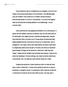
Continuing downwards, herniation of the umbilicus may present as an exomphalos or a protrusion of the gut as a gastroschisis, both which require extra caution in terms of infection control and urgent surgical repair to prevent gut herniation and strangulation. A gastroschisis has an incidence of 1:3000 with a tendency towards mothers under 20 and exomphalos comes with an incidence of 1:500 in mothers over 40 years of age. 5
Image 2: An example of a large exomphalos with a covering sac
Ending with the perigenital area and the limbs, common defects are imperforate openings of the anus, requiring surgical intervention to prevent bowel blockages and hypospadias in the male infant, where the urethra opens in the ventral or dorsal aspect of the penis rather than the glans. The limbs may show polydactyl (extra digits) or webbing between the digits of the hand or feet.
A heel prick test is commonly performed within the first week of birth. This tests for phenylketonuria, a disorder of phenylalanine metabolism that occurs in 1:10,000 births, leading to mental retardation if phenylalanine containing foods are fed and levels build up.6
Secondly, congenital hypothyroidism is also checked for, with an incidence of 3:10,000 babies. This leads to signs of infantile hypothyroidism, causing impaired physical and mental development.6
3.) What are the causes and implications of a diagnosis of foetal asphyxia?
Foetal asphyxia is a potentially serious diagnosis that requires rapid intervention by the obstetric team to prevent neonatal impairment or even foetal demise. Whilst foetal asphyxia may lead to organ ischaemia or even death, as in the case of an adult, it is not necessarily indicative of these outcomes, especially in early, mild cases.
Hypoxia, the initial stage of asphyxia develops when insufficient oxygenated maternal blood reaches the foetus. This can be due to a number of reasons, one being placental dysfunction. Problems such as placental artery insufficiency can lead to a decreased blood flow, likewise pre-eclampsia and maternal hypertension have the same effect. With pre-eclampsia, the increased oxygen demands of the mother in second stage labour leads to a diversion of blood away from the placenta and slight foetal hypoxia. The most severe placental cause is placental abruption, where the placenta shears away either completely or incompletely, leading to a severe fall in placental blood flow.
In this case, the cause of the foetal hypoxia and subsequent asphyxia is intrapartum. Normal transient drops in foetal heart rate are seen that correspond with uterine contractions and cord compression, leading to a fall in umbilical blood flow. A difficult, prolonged labour with poor progress increases the risk of foetal fatigue and loss of glycogen stores. This leads to the more ominous type 2 dips seen on cardiotocography, where the foetus does not recover its baseline heart rate as quickly as it should from the uterine contraction. A pathological cause is a twisted umbilical cord, either forming a few loops around the neck that can easily be clamped before the anterior shoulder is delivered, or many loops that can act as a noose as the baby is delivered, leading to a progressive loss of blood supply to the foetus. 3
The implications of foetal asphyxia vary with low hypoxia producing a jittery, hyperalert baby with hyperreflexia and generalised sympathetic activity, including tachycardia normal oculovestibular reflexes. The outcome is generally excellent, with 100% of all babies making a full recovery without any neurological deficit.
Moderate hypoxia produces a lethargic baby with overactive reflexes, increased bronchial secretions, seizures and bradycardia. The outcome is less positive, with 5% leading to foetal demise and 20% of those who survive developing neurological deficits.7
Severe hypoxia, as in this case, produces a stuporous, decerebrate posturing infant with poor reflexes, especially pupillary reflexes. The mortality rate ranges from 50-75%, with severe deficits in 80%, moderate deficits in 10% and normal children in 10%.3
- What is the clinical relevance of meconium staining and what are the risks associated with it?
Image 3: Image of a meconium stained baby.
Meconium consists of sloughed off intestinal epithelia, bile and mucus.8 It is the foetus’ first faeces to be developed. From Objective 3, I have described how mild hypoxia causes a generalised sympathetic drive increase, leading to foetal tachycardia and gut hypermotility. The anal sphincter is also relaxed in foetal hypoxia, leading to the expulsion of the gut contents into the amniotic liquor.
Around 9% of normal deliveries have meconium staining but suctioning of the airway is only indicated in an APGAR score of under five. The baby should be monitored in the first few hours of birth with a depression of vital signs indicating the need for NICU admission and a full workup.
The main risk to the neonate is if it inhales the meconium. Since the meconium contains gut digestive enzymes, the lung parenchyma is at risk for autodigestion leading to meconium aspiration syndrome.
Image 4: Meconium Aspiration Syndrome showing a left pneumothorax and interstitial damage.
Objective 4: What assisted vaginal delivery options are available and when are they indicated?
Assisted vaginal delivery techniques include forceps and ventouse methods. Thy are indicated in a delayed second stage leading to foetal distress or maternal exhaustion requiring the rapid delivery of the foetus. Other common causes of the need for operative delivery include cephalopelvic disproportion where the foetal head circumference is larger than the maternal pelvic outlet or the failure of the mother to push, for example in epidural anaesthesia the urge to bear down is commonly removed.
As this is an operative delivery, the obstetrician and the assistant should maintain sterile conditions including gowns and gloves, with the perineum cleaned and draped to prevent infection, as an episiostomy is usually required. The mother should be comfortable at rest and adequate anaesthesia given in the form of an epidural, spinal or sacral block as well as ensuring the cervix is dilated to 10 cms. For the foetus, no part of the cranium should be palpable in the abdomen and on internal examination the presenting part of the foetus should be at the ischial spine level, also known as position 0. Forceps are not used in breech delivery except to aid extraction of the head in a Caesarean section. 9
There are three main types of forceps in use today. These are the Wrigley’s, Simpson’s and Kielland’s forceps. The Wrigley’s is indicated when the presenting part of the foetus is at the level of the perineum and requires little effort in delivery whilst the Simpson’s forceps is used for deeper foetal heads as it has both a cephalic and pelvic curve, allowing the forceps to insert deeper. Both of these have a fixed, or English lock, allowing maximum traction to be applied. 10
In contrast, a Kielland’s forceps has a French sliding lock. This allows the two blades of the forceps to be offset against each other, allowing the babies’ head to be rotated in a certain direction.
Care must be taken when using a forceps to avoid damage to the foetal head but foetal bruising is common and non permanent, fading within 48 hours. More seriously, a facial palsy may be induced due to facial nerve compression; however this also is usually temporary. 9
For ventouse deliveries, a small suction cap is placed anteriorly to the occiput of the foetus and a vacuum of 0.8kg/cm2 is applied and the head is pulled with the line of the pelvis. Typically, a cephalohaematoma is produced that fades within the first week of life from the pressure of the vacuum. 10
Objective 6: What is the prognosis of this neonate?
This neonate initially had low APGAR scores at 1 minute and as a consequence, full resuscitation was initiated. He apparently suffered prolonged extreme hypoxia due to his low foetal heart rate, half of what is expected from a neonate.
His APGAR scores at 5 and 10 minutes were also extremely poor, indicating a very poor CNS outcome. This was confirmed by his grade 1 staging within 12 hours, progressing to a grade 3 stuporous state by 48 hours. The areas of infarction in both cerebral hemispheres and periventricular leucomalacia would suggest severe learning disorders and mental handicap, with the possibility of cerebral blindness and anosmia in later life. Cerebral damage is suggested by continuous generalised seizures that were unresponsive to medication.
Consequently, the prognosis is poor for this baby, with a high likelihood of a poor quality of life and a short life expectancy.
References
- Scottish Sensory Centre. Periventricular leucomalacia [online] [n.d.]. Available from: http://www.ssc.education.ed.ac.uk/resources/vi [accessed 12/3/2009].
- Tortora GJ, Grabowsky SR. Principles of anatomy and physiology. 10th ed. Hoboken, NJ : John Wiley and Sons, 2003.Hanretty KP.
- Obstetrics illustrated. 6th ed. Edinburgh : Churchill Livingstone, 2003.
- Nicholl R. What is the normal range of blood glucose in healthy term newborns? [online] [n.d.]. Available from: http://www.bestbets.org/bets/bet.php?id=252 [accessed 12/3/2009].
- Anaesthesia UK. Anaesthesia for neonates with abdominal wall defects [online] 2007. Available from: http://www.frca.co.uk/article.aspx?articleid=100983 [accessed 12/3/2009].
- Royal Free Hamstead NHS Trust (2004). The heel prick test [online] . Available from: http://www.royalfree.nhs.uk/pip_admin/Docs/heelpricktest_50.pdf [accessed 12/3/2009].
- Emedicine (2008). Hypoxic-Ischemic Encephalopathy [online] . Available from: http://emedicine.medscape.com/article/973501-overview [accessed 12/3/2009].
- Patient UK ([n.d.]). Meconium stained liquor [online] . Available from: http://www.patient.co.uk/showdoc/40000245/ [accessed 12/3/2009].Hamilton-Fairley D. Lecture notes:obstetrics and gynaecology. 2nd ed. Oxford : Blackwell, 2004.
- Symonds EM, Symonds IM. Essential obstetrics and gynaecology. 4th ed. Edinburgh : Churchill Livingstone, 2004.
- Hamilton-Fairley, J.A. (2004). Lecture notes: obstetrics and gynaecology. 2nd ed. Oxford : Blackwell.
Images
Image 1: http://www.bhj.org/journal/2002_4404_oct/images/fig2_700.jpg
Image 2:
Image 3:
Image 4: http://www.learningradiology.com/caseofweek/caseoftheweekpix/cow89.jpg