Discuss the Impact of Genome Sequences on the Study of Development
Cells and Development Discuss the Impact of Genome Sequences on the Study of Development Development refers to the biological process an organism undergoes during growth. The introduction of genetics this century has greatly accelerated our understanding in this field. It appears to be exponential, continually more scientists are being drawn into the field and more data is being generated. In this essay I will briefly outline the course of development as a subject over the past 100 years (with a slight bias towards animal development) commenting on how important the use of model organisms has become and the contribution to the field their genomes have made. Development started with Aristotle in the 4th century BC. He noted the different ways in which animals were born, oviparity, viviparity etc, and began to look at the transition from conception to adulthood. Not much happened in the study for about 2000 years, until a man named William Harves in 1651 made the profound statement that all animals are from eggs, "ex ovo omnia". The subject never really took off because the specimens were too small to analyse. The invention of the microscope revolutionised the science and allowed study of these once unseen structures. This coupled with the Morgan's' use of Mendel's' genetic theory to create the chromosomal theory of inheritance allowed scientists to begin to make

The arguments for and against developing a “genetic fingerprint” profile for all members of society
AS Module 4 Essay - The arguments for and against developing a "genetic fingerprint" profile for all members of society. Genetic fingerprinting is a rapidly developing technique involving the cutting of DNA and using it to distinguish between individuals of the same species. This is useful because every individual produces a unique genetic fingerprint as we all have different DNA sequences. Several steps are undertaken in order to prepare a genetic fingerprint. The non-coding DNA provides the basis of a genetic fingerprint (Potter, 2001). Firstly, a DNA sample is taken from, for example; the blood, a hair root or a mouth swab. If there is not enough DNA in the sample, a polymerase chain reaction (PCR) may be done to produce more. This is where the enzyme, DNA polymerase is used to amplify a piece of DNA by in vitro enymatic replication (http://en.wikipedia.org/wiki/Polymerase_chain_reaction, 02/03/2008). The next step would be to cut the DNA into pieces, and this is done by using the restriction endonuclease enzyme. The enzyme makes two incisions, one through each of the sugar-phosphate backbones (i.e., each strand) of the double helix without damaging the nitrogenous bases (http://en.wikipedia.org/wiki/Restriction_endonuclease, 02/03/2008). Now the DNA sections can be separated by the process of electrophoresis, which is a technique, used in the laboratories that result in
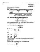
Determining Avogadro's Number Lab
Determining Avogadro's Number Lab Data Collection Table 1. Table showing initial mass, final mass and qualitative observation of copper electrodes. Electrode 1 Electrode 2 Initial Mass (g) ±0.005 g 5.77 5.80 Final Mass (g) ±0.005 g 6.27 5.27 Observations The copper electrode is shiny after being placed in copper sulfate. It also appears to have become thicker. The copper electrode is dull after being placed in copper sulfate. It also appears to have become thinner and rusty. Table 2. Table showing current of copper electrodes in 600 second intervals. Time (seconds) ±5 seconds Current (amps - A) ±0.05 A 0 .1 600 0.7 200 0.9 800 .0 Note: As both copper electrodes were placed in a single beaker of copper sulfate, the data obtained for the current is the same for both electrodes. Thus, the "Current" column in Table 2 represents the current of both copper electrodes. Table 3. Table showing voltage used during the experiment. Voltage (volts) ±0.5 V 6 Data Processing Moles: Number of moles Electrode 2, which lost 0.53 grams after being placed in copper sulfate, contains approximately 0.00834 moles of copper. Uncertainty of moles Uncertainty of mass = Uncertainty of initial mass + Uncertainty of final mass Uncertainty of mass = 0.005 g + 0.005 g Uncertainty of mass = 0.01 g Percent uncertainty of mass = Percent

How Zoo's Avoid Inbreeding in a Limited Captive Population
HOW CAN A ZOO WITH A LIMITED CAPTIVE POPULATION AVOID INBREEDING? * Contents. * Introduction. * Implications of Inbreeding in Limited Captive Population. * Solutions on Avoiding Inbreeding. * Ethical Issues How can Zoos with limited Captive Population Avoid Inbreeding Introduction; Animal Inbreeding was a problem in the early 1900's. London Zoo was one of the first and largest zoo's open in the world, Zoo keepers and scientists were ignorant and animals were kept in iron cages and confined spaces. They were not aware of the problem that inbreeding caused to captive populations. By the 1990's studies in plants and animals indicated that 'inbreeding depression' and many other effects were taking place naturally and in captivity. One solution to this was made in 1988 European zoos formed European Community Association of Zoos and Aquaria (EAZA), the countries involved were to keep detailed reports on animals which would be shared with other Zoos to keep breeding in the best possible interest of the animals. Implications of Inbreeding with limited Captive Population; Inbreeding is the term used when breeding within the family, close and distant in plants and animals, however, there are consequences of inbreeding, and bares illness and health problems within a population if continued over generations. Over generations more frequently recessive and deleterious traits can

Preperation of Antifebrin
Preparation of Antifebrin In this experiment, I am going to prepare the organic compound of antifebrin from readily available chemical reagents. Antifebrin is an odourless solid chemical of white flake-like appearance. Chemically, antifebrin is the amide phenylethanamide CH3ONHC6H5. It's slightly soluble in water. It does have the ability to self-ignite if it reaches the temperatures of 545°c but otherwise it's a stable compound. The pure crystals of antifebrin are plate shaped and white in colour. The antifebrin in this experiment is prepared from the reaction between phenylammonium chloride (C6H5NH3Cl) and ethanoic anhydride[ (CH3CO)2O ]. Chemical Equation for the Reaction: C6H5NH3+ Cl- + (CH3CO)2O CH3ONHC6H5 + CH3OOH + HCl Procedure & Observations: Procedure Observation Dissolve 1.0g of phenylammonium chloride in 30cm3 of water in a conical flask. Phenylammonium chloride is a greyish-green crystal like product. Adding water to it gives a solution pale grey with green tinge. After dissolving the solutions turns clear with a green-grey colour and no precipitate. Prepare a solution of 6.0g of sodium ethanoate in 25cm3 of water in a conical flask. Sodium ethanoate is a white powder. It dissolves completely in water to give a colourless solution. Carefully add 2cm3 of ethanoic anhydride to the solution of phenylammonium

An investigation into the effect of different sugars on respiration in yeast.
An investigation into the effect of different sugars on respiration in yeast. I am going to carry out an experiment, measuring the effect of different sugars on the respiration in yeast. In order to make a justified prediction I have researched different aspects of scientific knowledge, including respiration, yeast, sugar structure, enzymes and the collision theory. Glycolysis http://people.eku.edu/ritchisong/301notes1.htm Glycolysis is the splitting of a monosaccharide into two molecules of pyruvate. It takes part in the cytoplasm of a cell. Glycolysis begins with a monosaccharide with six carbon atoms, and ends with two molecules of pyruvate, each with three carbon atoms. For the first steps of glycolysis, energy from ATP is needed. However, energy is released in later steps to generate ATP. For every molecule of glucose, a net gain of two molecules of ATP is produced. The first stage of glycolysis is called phosphorylation, and results in hexose bisphosphate. This is shown in green on the above diagram. Hexose bisphosphate then breaks down into two molecules of triose phosphate. Hydrogen is removed from the triose phosphate and transferred to NAD to produce reduced NAD. These hydrogen's can then be used in oxidative phosphorylation to produce ATP. The end products of glycolysis are pyruvates, which still contains a lot of chemical potential energy. There are two

Chiral Drugs What is chirality? Chirality is the property possessed by a molecule with such spatial arrangement of atoms that it cannot superimpose on its mirror image.
Chiral Drugs What is chirality? "Chirality" is the property possessed by a molecule with such spatial arrangement of atoms that it cannot superimpose on its mirror image. The object and mirror- image pair of molecules has the same constituents and structural formula. Their relationship with each other is similar to our left and right hands. The carbon atom of a simple chiral centre has four different groups arranged tetrahedrally (Fig. 1). Isomers of such nature are called enantiomers. Fig.1: A chiral molecule with tetrahedral arrangement and its mirror image. There are three types of stereoisomers, namely enantiomers, diastereomers and geometrical isomers. . Enantiomers are two stereoisomers containing asymmetric carbon atoms related as non-superimposable object and mirror images. If an enantiomer rotates polarized light to the right or in a clockwise direction, it is said to be the (+) or the dextrorotatory isomer. On the other hand, if the plane polarized light is rotated to the left or in a counter-clockwise direction, the isomer is called as the (-) or the levorotatory isomer. Enantiomers are identical in chemical and physical properties except for the direction of rotation of plane polarized light. 2. Diastereomers are stereoisomers that are not related as object and mirror images. They contain at least two asymmetric carbon atoms. Unlike enantiomers, the

Determining the Water Potential of Sweet Potato Tissue
Determining the Water Potential of Sweet Potato Tissue Introduction The aim of this experiment is to determine the water potential of sweet potato tissue using osmosis. This can be achieved by placing the samples inside different molarities of sucrose solution and work out the percentage change in mass and then with the aid of a conversion graph convert molarity to water potential (kPa), without the weight of the sweet potatoes being a factor. Background Knowledge In mature plant cells, the fluid filled vacuole occupies most of the cell volume therefore in order to determine the water potential of the sample I would need to work out the water potential of this fluid inside the cell. Substances can pass in and out of cells by four different processes: * Diffusion * Osmosis * Active transport * Endocytosis & exocytosis All these processes involve substances passing through the cell membrane of the cell. In this investigation, I only need to consider osmosis. OSMOSIS is the movement of water molecules from a region of higher water potential to a region of lower water potential through a partially permeable membrane. This is a colligative property i.e. dependent on the concentration of particles in a solution. The water molecules involved always move down a ? gradient. It happens because of the natural kinetic energy possessed by the particles, which makes them move

Cellular Structure and Function
Cellular Structure and Function Introduction Cells are organised together into functioning groups called tissues. These groups of cells organise together to perform a specialised task. There are four basic types of tissues found in the human body; these are epithelial, connective, muscular, and nervous. The following text aims to explain the structure and function of these tissues as well as ovum and sperm cells. Epithelial Tissue Epithelial tissues, as with all different types of tissue, can be found all over the body, they generally line the inside or outside of a body cavity. The cells are anchored down by basement membranes and form in different shapes such a flat, cuboidal, and columnar. The below text details several different types of epithelium cells that can be located around the body. . Stratified Squamous Epithelium Stratified squamous epithelium cells are present in areas of the body that are very moist and subject to abrasion, such as the mouth, esophagus and vagina. The cells are packed densely together and are very flat and irregular in shape. The tissue functions to provide a barrier to entry to inside the body and protects underlying tissues from friction and drying. Figure A. above shows a drawing of the epithelia from an overhead point of view, in shape they are very similar to that of a fried egg. Figure B. shows the cells from a side-on point of

Cellular organelles Structure and Function
Eukaryotic Cellular Organelles: Structure and Function Introduction Cells take many different forms in living organism’s but there are a certain common features in which eukaryotic cells share. Individual cells consist of minute membrane bound vesicles found in the cytoplasm called organelles. These organelles play a crucial role in undertaking the processes that go in inside the cell in order for it to function. The following text aims to explain the structure and function of the major organelles. Nucleus The nucleus is commonly referred to as the control centre or brain of the cell; it directs instructions to other organelle to carry out specific tasks and contains the genetic material of the cell. The long strands of DNA found in the nucleus combine with proteins to form chromatin; the chromatin is then used to create chromosomes. A: Chromatin B: Nuclear Pores C: Nuclear Envelope D: Nucleolus E: Chromosomes Above: A Nucleus. The Nucleus is enclosed entirely by an inner and outer nuclear membrane which protects the fragile DNA and genetic material within. Throughout the surface of the nucleus the two membranes fuse together to create pores that allow the exit of RNA, and the entrance of nucleotides for DNA replication. Endoplasmic reticulum (ER) The endoplasmic reticulum is a network of folded tubules and vesicles found on the outside of the nucleus. Part of


