Enzymes Revision notes.
Introduction Enzymes are biological catalysts. Many chemical reactions can be speeded up by substances called catalysts. A catalyst alters the rate of a chemical reaction , without taking part in it itself. In Living organisms chemical reactions take place all the time. Almost all of these are controlled by catalysts called enzymes. For example, in the alimentary canal, large molecules are broken down to smaller ones through digestion. these are sped up by enzymes. a different enzyme is needed for each kind of food. E.g. starch is digested to the sugar maltose by an enzyme called amylase. Protein is digested to amino acids by protease. Each enzyme can be used over and over again. this means that a small amount of enzyme can catalyse the conversion of a lot of substrate into a lot of product. Enzymes have Active sites Enzymes are proteins. Their molecules have very precise 3 dimensional shapes. their shame include a dent , which is exactly the right size and shape for a molecule of the enzymes substrate to fit into . this dent is called the active site. When a substrate molecule slots into the active site , the enzyme tweaks the substrate molecule , pulling it out of shape and making it split into the new products molecules. This is sometimes called the lock and key method. the enzymes active site is the lock and the substrate is the key . only the correct key will fit

Cell Structure
Cell Structure Difference between resolution and magnification Magnification Resolution Magnification is the factor by which an image is enlarged. Resolution is the smallest distance that can be between two objects so that they still appear distinctly separate in the microscope picture. Determined by the size and magnitude of the lenses used Determind by the wavelength of light Difference between light microscope and electron microscope Light Microscope Electron microscope Uses light source focused by transparent lenses Uses electrons focused by magnetic lenses Lower resolution(usually 0.2 micrometer) Higher resolution(usually 2 nanometer) Allows study of living cells Doesn't allow study of living cells Specimens are stained with colourful dyes such as iodine Specimens are stained with heavy metal ions such as lead Specimens can be relatively thick Specimens are to be very thin because of less penetrating power of electrons Cell Organelles Eukaryotic cells Organelle Main function Structure Organisms Notes chloroplast (plastid) * Contains molecules for the light dependant stage of photosynthesis * Synthesis of ATP * Enzymes required for conversion of CO2 and water to carbohydrate * Temporary storage of carbohydrate Sacs called thylakoids formed by membranes Stroma contains enzymes plants, protists (rare kleptoplastic organisms) has some
The Heart, Structure and Function
Heart Structure and Function Mammals have a double circulation Pulmonary Circulation- Takes blood on the relatively short return journey to the lungs, where blood is oxygenated Systemic Circulation- Takes blood around the rest of the body The human heart is covered by a double layer of tough inelastic membranes which form the pericardium. A fluid (pericardial fluid) is secreted between the membranes, allowing them to move easily over each other. The pericardium protects the heart from over-expansion caused by elastic recoil when it is beating very fast. The walls of the heart consist mainly of cardiac muscle, a special type of muscle only found in the heart. Unlike other muscles, it never fatigues. However it does not tolerate lack of oxygen or nutrients and soon dies if its supply of blood is cut off. The heart is divided into a left side and a right side by the septum. The septum becomes rigid just before the heart contracts, so that it functions as a fulcrum for the action of the heart muscle. Each side of the heart has 2 chambers: an atrium which receives blood from the veins and a ventricle which pumps blood into arteries. Deoxygenated blood from the systemic veins enter the right atrium and is passed through the tricuspid valve into the right ventricle. This contracts and pumps blood through the pulmonary artery into the lungs. Oxygenated blood returns through
Testing the tensile strength of celery
Tensile Strength of Celery Stems Experiment This experiment aims to measure the tensile strength of fibres from the stem of the celery plant. Celery is an herbaceous plant grown for both eating and as a medicinal. Aim The aim of this investigation is to measure the tensile strength of celery fibres. Hypothesis I believe that the tensile strength of the celery fibres will very much depend on how thick and healthy the sample is. I reckon that the celery will have a high tensile strength, however it is difficult to investigate 100% fairly. Apparatus * Celery * Knife * Clamp stand and clamp * Sticky tape * Weights and Weight holder. Method First of all, we were unable to do a control experiment with the celery because we did not have the apparatus. However, if we did, one possible method would be to damage the fibres by heating them, perhaps by placing them in boiling water for a few minutes. We would then use the following method. Initially, we cut the stem with a knife and gently peeled away fibres with the help of a mounted needle. Next, we needed to attach the fibre to the clamp stand in order to measure its strength. However, this proved to be difficult to do effectively. After a bit of initiation, we fixed the fibre to the stand by using sticky tape to hold it in place. We then added weights 10g at a time being careful not to drop the weights suddenly as
Adaptations of Small Intestine for Absorption
MARIS STELLA HIGH SCHOOL SEC 3 PURE BIOLOGY Topic 6 Nutrition in Mammals T6.3 Absorption of Food & Functions of the Liver . Adaptations of Small Intestine for Absorption It is very LONG, about 5-6 m. This gives plenty of time for digestion to be completed, and fordigested food to be absorbed as it passes through. It has very FOLDED surface. The inner wall is thrown into numerous folds & furrows. Thus the surface area for absorption is greatly enlarged. It has minute finger-like projections (about 1 mm long) called villi covering the whole internal surface of the small intestine. (Each square millimemtre of surface has about 20-40 villi and there are about 5 million in the ileum.) Each villus bears even smaller projections, microvilli. The villi & microvilli further increase surface area for absorption. Villi contain network of blood capillaries & lacteals for transporting digested, absorbed food away. One-cell thick epithelium. The digested food can easily cross the wall to reach the capillaries & lacteals. *There are intestinal glands between bases of villi. These secrete intestinal juice to help in digestion. (refer to T6.2) 2. Summary of Absorption 1. Simple sugars, amino acids, vitamins & minerals l (diffusion) blood capillaries of villi l hepatic portal vein l liver --- hepatic vein --- heart 2a. Soluble glycerol l (diffusion) epithelium

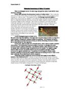
The transport system in plants moves water soluble molecules by vascular tissue. There are two types of vascular tissue which are Xylem and Phloem.
Xylem and Phloem The transport system in plants moves water soluble molecules by vascular tissue. There are two types of vascular tissue which are Xylem and Phloem. Both of them are specialised to carry out their role in the plant and they are normally found together in vascular bundles throughout the plant. The xylem tissue transports water and soluble minerals up the plant towards the leaves for photosynthesis whereas the phloem tissue transports the sugars made by the reaction up and down the plant to places where it is needed. Both of the tissues are highly specialised and they have other tissues accompanying them in the vascular bundles to give strength. Most importantly the tissues do not use any energy or transport mechanism to move the water based molecules however the water or water based substance itself has cohesive properties. The cohesive properties of water cause molecules to attract to each other and the surface tension in the vessels keeps it moving up towards the leaves. As the water on the leaves is evaporating or being used in a process it causes water to move up the plant and more water to be taken in by the root's which is also seen as an ongoing cycle. Xylem The xylems are many endless vessels starting from the roots and going to all the area of a plant. The walls of the vessel are made up of dead cells strengthened by lignin forming an endless tube.
Core PracticalDoes caffeine affect heart rate?)
Core Practical Does caffeine affect heart rate? Introduction: -: Caffeine is the name of an alkaloid present in plants, such as coffee and tea.Caffeine is absorbed rapidly into the bloodstream from the gastro-intestinal tract. Throughout the body it increases metabolic rate by around 10%. Caffeine has many metabolic effects. For example, * It stimulates the central nervous system * It releases free fatty acids from adipose (fatty) tissue * It affects the kidneys, increasing urination, which can lead to dehydration * Increase heart beat Hypothesis: - My hypothesis was that the heartbeat of the water flea would increase when placed in a caffeine solution. Method:- * We took a cavity slide and placed some cotton wool on it and then placed a water flea on to the cotton wool two drops of water were added with the help of a pipette. * The cavity slide was placed under a microscope. We used two stop clocks to measure heartbeat of water flea in periods of 15 sec /min. We took five heartbeat readings. * This method was repeated six times each time a solution of caffeine was added. First we added .2%caffieine and placed a filter paper to stop the water evaporating. On the last occasion amount of caffeine that was used was 0.5% solution. No of heartbeats in 15 sec/min .RESULT:- Length of treatment(min) Time(min) 2 4 6 8 10 Treatment Control 43/172 61/244
Blood structure and function in the body.
Blood Blood can be defined as: the circulating fluid (plasma) and suspended formed elements, such as red blood cells, white blood cells and platelets in the vascular system of humans and other vertebrates. This essay will mention about structure of the blood and the functions of the blood. Structure of blood has four major parts these are: plasma, red blood cells, white blood cells, and platelets. Plasma is mainly made up from water; nearly %90-%92 is water. This is a straw coloured fluid. Plasma contains dissolved substances including electrolytes for example: sodium, chlorine and potassium. Albumin, globulin and fibrinogen are the protein of blood plasma. Hormones are also in the blood plasma. Red blood cells also named as erythrocytes. Red blood cells sometimes may be immature and they do have nucleus but in general, the mature ones do not have any nucleus. This gives them ability to carry more oxygen. They only exist for 120 days in body and there are approximately 4.5 - 5.8 million erythrocytes per micro-litre of healthy blood. This quantity depends on gender and age. White blood cells also named as leucocytes. There are different types of leucocytes. These can be defined as; granular and agranural. Depending on their types, they sometimes exist for few hours to few days but sometimes they can exist for many years. Platelets also called as trombocytes. These are
The Biological Importance of Water
Biological Importance of Water (10 marks) Water is a biological marvel. Its wide range of properties makes it essential for many reactions in Biology. Firstly, water produces the phenomenon known as 'surface tension'. This is when the molecules of water at the surface are held together making a surface strong enough for some organisms to 'walk on water'. This happens because the hydrogen atoms are slightly ?+ whereas the oxygen atoms are slightly ?-. Lots of these water molecules together mean that there is a slight intermolecular attraction, because the slightly negative oxygen is attracted towards the slightly positive hydrogen. This results in a hydrogen or H-bond, although technically speaking it is not actually a bond, just an attraction. As these molecules move around, the H-bonds continuously break and reform with other molecules. Although individually, these bonds are weak, the collective action of breaking and reforming means that together, the force is very strong. An example of an organism relying on the concept of surface tension is the basilisk lizard, also known as the Jesus Christ lizard because of its ability to race across the surface of water: Another biological property of water is the fact that it has a high heat capacity. This means that it requires a large increase in energy for a small increase in temperature, making it brilliant for sweating. This
Observing Mitosis. The purpose of this experiment is to prepare a slide of actively dividing plant tissue and to observe stages of the cell cycle in living tissue.
Observing Mitosis The purpose of this experiment is to prepare a slide of actively dividing plant tissue and to observe stages of the cell cycle in living tissue. We had to consider the duration of mitosis in relation to the whole cell cycle. First of all got some hydrochloric acid and put it into a water bath at 60°C. Then we cut off some root tips from garlic, we used the tips because it's where mitosis still takes place - the meristem. We put these roots into some acetic alcohol which is a fixative, so it stops and helps preserve the cells. Then we removed them and put them in ice cold water and then dried them on filter paper. After this we put the root tips in the pre-heated hydrochloric acid, this will dissolve the calcium pectate but leave the cell walls unharmed. So it breaks up tissue without damaging the cell. After cooling down again and drying, we put the root tips onto a clean microscope slide and cut off the tip of it. We had to make sure it was the meristem and not other differentiated root tissue. Then we macerated the root tip, this breaks up all the cells and makes them one layer. If not sufficiently macerated the layer may not be thin enough to observe cell nuclei. After this we added a small drop of acetic orcein stain so that we could see the cells under the microscope. We then added a cover slip over the cells, and blotted firmly to get rid of