
Too Much Information: Genetic Testing
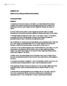
Too Much Information: Genetic Testing Biology OAC ISP Essay - By Daniel Perez Genetic testing offers a whole new world of information about us and how our bodies work. The data we get from delving into our own genetic code can help us to cure or even prevent disease, stop medical conditions such as cancer or cystic fibrosis from even manifesting, or even correct these sorts of errors before birth, and many other beneficial uses. However, at this point in time, all of this is beyond us. We have no miracle cures, no 'magic bullet' with which to fight disease or genetic conditions, in fact, our understanding of the genetic code is so limited that it's as if we cannot see the forest for the trees. We have taken our first baby steps into understanding human genetics with the completion of the Human Genome Project, and now that we have the big picture, we can begin to interpret it. Through information gleaned from our DNA, we now know that there are certain medical conditions that are caused by certain patterns within the genes. Some examples of these genetic conditions include Tay-Sach's disease, Bloom syndrome, Deafness, cystic fibrosis, and many other diseases (http://www.einstein.edu/e3front.dll?durki=7158). Although many of these conditions are fatal, the ones that are not can be treated early, even before symptoms develop when possible, or if not treated, at least monitored
Titration Lab Report
CHEMISTRY LAB Titration Curves of Strong and Weak Acids and Bases Processing the Data: Questions: . Examine the time data for each of the Trials 1-4. In which trial(s) did the indicator change color at about the same time as the large increase in pH occurred at the equivalence point? In which trial(s) was there a significant difference in these two times? In all the 4 trials, the time taken for color change and the time taken for a large increase in pH was the same, leaving no significant difference between the two values. 2. Phenolphthalein changes from clear to red at a pH value of about 9. According to your results, with which combination(s) of strong or weak acids and bases can phenolphthalein be used to determine the equivalence point? The combination of a Strong Acid and Base will give us the equivalence point: there will be a color change of phenolphthalein at pH 9. It is also observed that the reaction between a Weak Acid and Strong Base can be used to obtain a pH of 9. 3. On each of the four printed graphs, draw a horizontal line from a pH value of 9 on the vertical axis to its intersection with the titration curve. In which trial(s) does this line intersect the nearly vertical region of the curve? In which trial(s) does this line miss the nearly vertical region of the curve? For Trials 1 and 3, the horizontal line from pH 9 intersects the S curve. For

The advantages and limitations of electron microscopy.
The advantages and limitations of electron microscopy There are two main branches of microscopy that are pertinent to cell biology. These branches arise from the two types of microscope; the light microscope and the electron microscope. The basic principles of light microscopy have been known since circa 17th century, however improvements in lens manufacture in circa 19th century allowed the use of microscopy to be much more practically available and useful. This is increased ability inspired rapid research into both the design of microscopes and the preparation of specimens. However, the light microscope can only magnify objects bigger than 0.2 micrometres; due to its limited resolving powers. This is because it utilises a beam of light. Relatively, light has a long wavelength, this means that when there are two small points close together there is too much refraction and wave front overlap, the eye then only sees one point. This can also be considered in terms of objects "crossing the path" of the wavelength. The smallest wavelength of visible light is 400nm, the diameter of mitochondria is 1000nm, and therefore mitochondria cross the path of the light wave. However ribosomes have a diameter of 22nm, and do not cross the path of the light wave and are therefore not seen by the light microscope. As biologists came to realise these limitations they understood that the

investigating the relationship between the diameter and the current in a wire at its melting point
Investigation Report Aim Theory Electrical resistance is a measure of the degree to which an object opposes the passage of an electric current. The SI unit of electrical resistance is the ohm. Its reciprocal quantity is electrical conductance measured in siemens. Resistance is the property of any object or substance of resisting or opposing the flow of an electrical current. The quantity of resistance in an electric circuit determines the amount of current flowing in the circuit for any given voltage applied to the circuit. Some formulae for resistance are where R is the resistance of the object / ? V is the potential difference across the object / V I is the current passing through the object / A (Ref. http://en.wikipedia.org/wiki/Electrical_resistance) where R is the resistance/ ? ? is the resistivity / ?m l is the length of the wire / m A is the cross section area of the wire / m A = ?() = ? where A is area / m d is the diameter / m Putting the formulae together, so (Ref. http://physics.bu.edu/~duffy/PY106/Resistance.html) Aim of investigation The aim of this work is to investigate the relationship between the resistance and the diameter of the wire. Variables Variable Independent / Controlled / Dependent Resistance D Resistivity C Length of wire C Diameter I Prediction Since the theory suggests that So So the resistance should be
Cystic Fibrosis
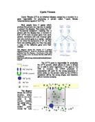
Cystic Fibrosis Cystic Fibrosis (CF) is an inherited disease caused by a mutation in a gene responsible for producing a protein called "cystic fibrosis transmembrane regulator" (CFTR). Most people have 2 genes which produce this protein, but only one is needed to prevent the disease. This means that CF is "autosomal recessive", meaning that a person with the disease has a mutation in both CFTR (one mutated gene from each parent). Someone with one mutated gene and one normal gene is a carrier. Carriers do not show the symptoms of CF, as they have one working gene, but they may pass a copy of the defective gene onto their children. The CFTR gene is responsible for producing the CFTR protein, which allows Cl- ions to diffuse out of cells in water regulation. If the gene in the DNA is mutated, the mRNA produced in transcription will code for the wrong sequence of amino acids, so the protein made by the mRNA in translation will be the wrong shape, and therefore will not function correctly. This diagram shows the normal situation, where there is too much water in the mucus (outside the apical end of the cell). The sodium pump moves Na+ ions out of the cell, into the tissue fluid outside the basal end of the cell. The Na+ channel allows sodium ions to diffuse into the cell to replace those lost, causing a more negative water potential in the cell, so water moves out of the mucus

Investigating the breakdown of hydrogen peroxide using celery tissue to supply the enzyme catalyst
Investigating the breakdown of hydrogen peroxide using celery tissue to supply the enzyme catalyst Variables * Amount of celery * Concentration of celery, more or less watered down. * Concentration of Hydrogen Peroxide (H202) * The amount of H202 * The temperature of H202 I am going to vary the concentration of the hydrogen peroxide. I think that varying the concentration of the liquid will be the best experiment to do and will hopefully give a strong set of results, which will enable me to obtain clear conclusions. Prediction The rate of an enzyme- controlled reaction depends on the temperature, pH, and concentrations of the enzyme and its substrate. The more enzyme molecules produced by a cell, the faster the reaction will proceed. Similarly, an increase in the substrate concentration will speed up the reaction if there are enough enzymes molecules to cope with the additional substrate. Therefore by diluting the hydrogen peroxide with water, this will decrease the rate of decomposition of the H202, and the less gas will be given off. The enzyme in the experiment is catalase. Hydrogen peroxide is poisonous and the catalase works to render the hydrogen peroxide harmless by breaking it down to water and oxygen. If the concentration of H202 is less, then there is more water present, and there are less hydrogen peroxide molecules, so there is less for the catalase to
Investigate the effect of pH on Trypsin
Biology Coursework Plan Aim Investigate the effect of pH on Trypsin Prediction / hypothesis * pH will affect trypsin action * as pH increases, tryrpsin will show increasing activity up to an optimum pH * the action of the enzyme trypsin on the substrate egg albumen will be at a maximum at an optimum pH of around 7 (neutral) to 8 (slightly alkaline) see later about pH of duodenum * as pH continues above this, trypsin activity will decrease Background / Introduction Proteins are complex organic compounds consisting of amino acids joined by peptide bonds which form highly folded three dimensional or tertiary structures. The bonds that maintain the tertiary structure of the protein are a result of interactions between the R groups of the amino acids: disulphide bridges (strong covalent), hydrogen (weak) bonds, ionic or electrovalent bonds, hydrophobic interactions. Enzymes, such as trypsin, are globular proteins with a specific shaped active site into which the correct substrate can fit. trypsin protein / polypeptide peptides Trypsin is a protease enzyme: a hydrolytic or digestive protein that cleaves peptide bonds. It is produced in the pancreas in the form of trypsinogen, and is then transported to the duodenum of the small intestine, where the digestion of proteins to polypeptides and amino acids begins. The pH

Cellular Respiration and the Role of Mitochondria
Cellular Respiration and the Role of Mitochondria Cellular respiration is the process of oxidising food molecules, such as glucose, to carbon dioxide and water and releasing the covalent bond energy in the form of ATP for use by all the energy-consuming activities of the cell. Mitochondria are membrane-enclosed organelles distributed through the cytosol of most eukaryotic cells. They are where cellular aerobic respiration occurs; indeed cells without mitochondria cannot respire aerobically. Cellular respiration consists of two broad phases, initially, glycolysis (the breakdown of glucose to pyruvic acid) Occurs, this is followed by the oxidation of pyruvic acid to carbon dioxide and water. In eukaryotes, glycolysis occurs in the cytosol (The fluid in which cell organelles are suspended). The remaining processes take place in the mitochondria. The first stage, glycolysis is the anaerobic catabolism of glucose, it occurs in almost all cells. The process uses glucose and co-enzyme NAD (Nicotinamide Adenine Dinucleotide), and yields 2 molecules of Pyruvic acid, as below C6H12O6 + 2NAD+ -> 2C3H4O3 + 2NADH + 2H+ The free energy stored in 2 molecules of pyruvic acid is somewhat less than that in the original glucose molecule. Some of this difference is captured in 2 molecules of ATP. The Krebs Cycle then decarboxylates the pyruvic acid resulting in a 2-carbon fragment of
The structure and function of the ileum in relation to absorption and digestion.
SUMUDU LANKATILAKE 7-FEB-03 THE STRUCTURE AND FUNCTION OF THE ILEUM IN RELATION TO ABSORPTION AND DIGESTION The ileum is the second part of the small intestine located after the duodenum. It has a vital function in digestion and has a suitable structure to accommodate for its functions. The ileum is 6 meters long and the main site foe the absorption of the soluble products of digestion. The ileum is efficient at this for the following reasons: * It is fairly long and presents a large absorbing suface to the digested food. * Its internal surface is greatly increased by circular folds bearing thousands of projections called villi. These villi are about 0.56mm long and may be finger like or flattened in shape. * The lining epithelium is very thin and the fluids can pass rapidly through it. The outer membrane of each epithelial cell has microvilli which increase the exposed surface of the cell by 20 times. * There is a dense network of blood capillaries in each villus for quick absorption and maintainance of the concentration of the concentration gradient. * The villi possess smooth muscle fibres that contract and relax and mix the food up and bring it into contact with the epithelial cells of the absorptive surface. * Each villus has a lacteal for the absorption of fatty acids and glycerol, most of which combine to form fats. The ileum is made up of 4 layers:

structural differences between fibrous and globular proteins.
Question: Explain with examples, the structural differences between fibrous and globular proteins. A globular protein has a fixed specific sequence of amino acids that are non-repetitive while a fibrous protein has a repetitive regular sequence of amino acid. For example, haemoglobin, a globular protein is made up of 4 polypeptide chains to form a tetramer (?2?2), composed of two identical alpha-beta (??) dimers. Collagen, a fibrous protein, has a primary structure characterized by a repeating tripeptide sequence of Glycine - X - Y. (X is proline, Y is either hydroxyproline or hydroxylysine) A globular protein has a more compact structure owing to highly contorted pattern of folding, bending and twisting along polypeptide chain to give the protein a spherical 3D shape while a fibrous protein is usually formed with elongated polypeptide chains wrapped around to form multi-molecular paralleled filaments to strands. For example, haemoglobin is a tetramer made up of 4 polypeptide chains of 2? chains and 2? chains. These four subunits are packed to form an overall spherically shaped molecule. However, collagen, a fibrous protein, is formed with three polypeptide chains lie parallel and wind round one another, forming a tropocollagen. The tropocollagen molecules lie side by side and are linked to each other giving a collagen fibril. A globular protein has its length of


